Licensing

Pd
Public Domain: This image is in the public domain and thus free of any copyright restrictions. However, as is the norm in scientific publishing and as a matter of courtesy, any user should credit the content provider for any public or private use of this image whenever possible. Learn more
Comments: Powered by Pnyxe

Description

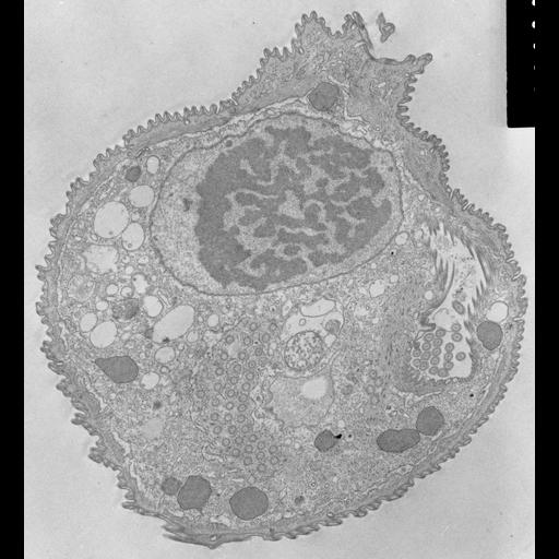
This figure shows the connection between the peristome and the food vacuole forming zone where the alveoli end. The cytostome is the opening that connects these two zones. TEM taken on 6/13/69 by R. Allen with Philips 300 operating at 60kV. Neg. 6,370X. Bar = 1µm. A print of the negative was scanned and processed in Photoshop. The raw negative was scanned with an Epson Perfection V750 Pro and this high resolution image is best used for quantitative analysis. Additional information available at (http://www5.pbrc.hawaii.edu/allen/).

Technical Details

Standard glutaraldehyde fixation followed by osmium tetroxide, dehydrated in alcohol and embedded in an epoxy resin. Microtome sections prepared at approximately 75nm thickness.

Biological Sources

NCBI Organism Classification
Opercularia [NCBITaxon:168247]
Opercularia coarctata
Cell Type
cell by organism
eukaryotic cell
Eukaryotic Protist
Ciliated Protist
Cell Line
Cambridge Culture Collection, Cambridge, England, # LB1657/1
Cellular Component
macronucleus
oral apparatus
cell cortex
cytoplasm
cytostome

Attribution

Name
Richard Allen
Link
http://www5.pbrc.hawaii.edu/allen/

Grouping

This image is part of a group.
Data Qualifiers
raw, unprocessed data
suitable for spatial measurements

Dimensions

Spatial Axis Image Size Pixel Size
X 3651px 1.6nm
Y 4000px 1.6nm
*CIL – Cell Image Library accession number. Please use this to reference an image.