Alternate header for print version


Licensing
Attribution Non-Commercial Share Alike:This image is licensed under a Creative Commons Attribution, Non-Commercial Share Alike License. View License Deed | View Legal Code
tweet  
*CIL – Cell Image Library accession number. Please use this to reference an image.

CIL:39326*  Cite 
Description

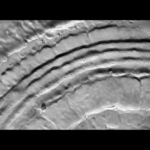
This freeze fracture view of the pellicle of V. microstoma near the aboral end of the zooid shows the region where the width of each circumferential alveolus is narrower and thus the strings of fractured pits are closer together. In this case the E-fracture face of the outer alveolar membrane is shown which has fewer IMPs than the P-face of the outer alveolar membrane. Here the connections are indentations rather than fractured necks. In a few areas the P-fracture face of the plasma membrane is viewed where it was pinched into the folds between rings that surround the cell. The plasma membrane demonstrates the large number of prominent IMPs on this face of the plasma membrane. TEM taken on 11/11/75 by R. Allen with JEM 100B. Neg. 21,578X. The raw negative was scanned with an Epson Perfection V750 Pro and this high resolution image is best used for quantitative analysis. Additional information available at (http://www5.pbrc.hawaii.edu/allen/).

Biological Sources
NCBI Organism Classification
Vorticella microstoma
Cell Type
cell by organism
eukaryotic cell
Eukaryotic Protist
Ciliated Protist
Cellular Component
plasma membrane
cell cortex
cell cortex part
pellicle
Biological Context
Biological Process
plasma membrane organization
Attribution
Names
Richard Allen (University of Hawaii)
Citation
Digital Object Identifier (DOI)
doi:10.7295/W9CIL39326
Archival Resource Key (ARK)
ark:/b7295/w9cil39326
Grouping This image is part of a group.
Imaging
Image Type
transmission electron microscopy (TEM)
illumination by electrons
Image Mode
electron density
Parameters Imaged
elastic scattering of electrons
electron density
elevation
Source of Contrast
differences in deposition of metal shadow
Visualization Methods
transmission electron microscopy (TEM)
Processing History
freeze_fracture/freeze_etch
shadowing and plating
recorded image
film
Data Qualifiers
raw, unprocessed data
suitable for spatial measurements
Dimensions
Spatial Axis Image Size Pixel Size
X 4543px 0.93nm
Y 3327px 0.93nm