Licensing

Pd
Public Domain: This image is in the public domain and thus free of any copyright restrictions. However, as is the norm in scientific publishing and as a matter of courtesy, any user should credit the content provider for any public or private use of this image whenever possible. Learn more
Comments: Powered by Pnyxe

Description

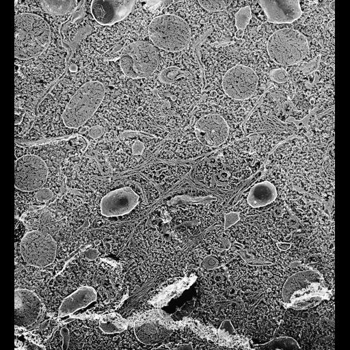
The infraciliary lattice in this quick-freeze deep-etched image consists of a meshwork of branching fibers composed of many subfibers. Basal bodies and trichocyst tips extend through the openings of the mesh. Individual metal-coated fibrils of the mesh average 11 nm in diameter and are closely interlaced and cross-linked to other fibrils. TEM taken on 5/27/92 by R. Allen with Zeiss 10A operating at 80kV. Neg. 9,780X. The raw negative was scanned with an Epson Perfection V750 Pro and this high resolution image is best used for quantitative analysis. Additional information available at (http://www5.pbrc.hawaii.edu/allen/).

Biological Sources

NCBI Organism Classification
Paramecium multimicronucleatum
Cell Type
cell by organism
eukaryotic cell
Eukaryotic Protist
Ciliated Protist
Cellular Component
cell cortex
mitochondria
trichocyst
cilium
cytoplasm

Biological Context

Biological Process
cortical cytoskeleton organization
cytoplasm organization

Attribution

Name
Richard Allen (University of Hawaii)
Link
http://www5.pbrc.hawaii.edu/allen/

Grouping

This image is part of a group.
Data Qualifiers
raw, unprocessed data
suitable for spatial measurements

Dimensions

Spatial Axis Image Size Pixel Size
X 5498px 1.4nm
Y 6001px 1.4nm
*CIL – Cell Image Library accession number. Please use this to reference an image.