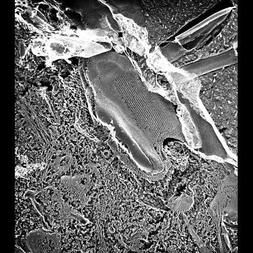
In quick-freeze deep-etch preparations the plasma membrane can be seen to contain plaques or plates of intramembrane particles (IMPs) that are regularly aligned in rows with a center-to-center spacing between rows of 25nm. These plaques remain in the P-fracture face of the plasma membrane and are found mostly on the anterior ventral surface of the cell near the anterior suture. Their function is not known. For discussion see Allen, J. Ultrastruc. Res. 63:64-78, 1978 and Hufnagel, Micros. Res. Tech. 22:225-264, 1992. TEM taken on 5/26/92 by R. Allen with Zeiss 10A operating at 80kV. Neg. 19,800X. The raw negative was scanned with an Epson Perfection V750 Pro and this high resolution image is best used for quantitative analysis. Additional information available at (http://www5.pbrc.hawaii.edu/allen/).
| Spatial Axis | Image Size | Pixel Size |
|---|---|---|
| X | 4892px | 0.8nm |
| Y | 5550px | 0.8nm |