Alternate header for print version


Licensing
Attribution Non-Commercial Share Alike:This image is licensed under a Creative Commons Attribution, Non-Commercial Share Alike License. View License Deed | View Legal Code
tweet  
*CIL – Cell Image Library accession number. Please use this to reference an image.

CIL:39306*  Cite 
Description

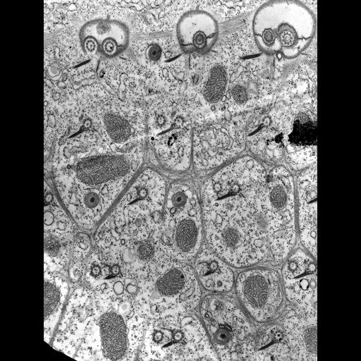
This EM features the infraciliary lattice which forms a continuous network at the level of the proximal ends of basal bodies where the kinetodesmal fibers are connected to the posterior basal bodies of a pair. This system is separated by a small gap from the more superficially located striated bands. The gap has no apparent fibers connecting these two systems. This network forms a highly porous boundary between the ectoplasm and the endoplasm. Electron dense posts are located at each branching site of this network. It is now known that this network is composed of centrin, a contractile protein also found in centrosomes of higher animals including man. TEM taken on 12/18/68 by R. Allen with Philips 300. Neg. 16,000X. Published in J. Cell Biol. 49:1-20, 1971.

Technical Details

Standard glutaraldehyde fixation followed by osmium tetroxide, dehydrated in alcohol and embedded in an epoxy resin. Microtome sections prepared at approximately 75nm thickness. Additional information available at (http://www5.pbrc.hawaii.edu/allen/).

Biological Sources
NCBI Organism Classification
Paramecium multimicronucleatum
Cell Type
cell by organism
eukaryotic cell
Eukaryotic Protist
Ciliated Protist
Cellular Component
plasma membrane
cell cortex
Biological Context
Biological Process
cortical cytoskeleton organization
Attribution
Names
Richard Allen (University of Hawaii)
Published
J Cell Biol. 1971 Apr;49(1):1-20.
Pubmed
4102004
Citation
Digital Object Identifier (DOI)
doi:10.7295/W9CIL39306
Archival Resource Key (ARK)
ark:/b7295/w9cil39306
Imaging
Image Type
transmission electron microscopy (TEM)
illumination by electrons
Image Mode
detection of electrons
film
Parameters Imaged
transmission electron microscopy (TEM)
Source of Contrast
electron density
stain with broad specificity
Visualization Methods
stain with broad specificity
osmium tetroxide
uranyl salt
lead salt
Processing History
recorded image
film
Data Qualifiers
raw, unprocessed data
suitable for spatial measurements
Dimensions
Spatial Axis Image Size Pixel Size
X 4772px 0.94nm
Y 6288px 0.94nm