Alternate header for print version


Licensing
Attribution Non-Commercial Share Alike:This image is licensed under a Creative Commons Attribution, Non-Commercial Share Alike License. View License Deed | View Legal Code
tweet  
*CIL – Cell Image Library accession number. Please use this to reference an image.

CIL:39168*  Cite 
Description

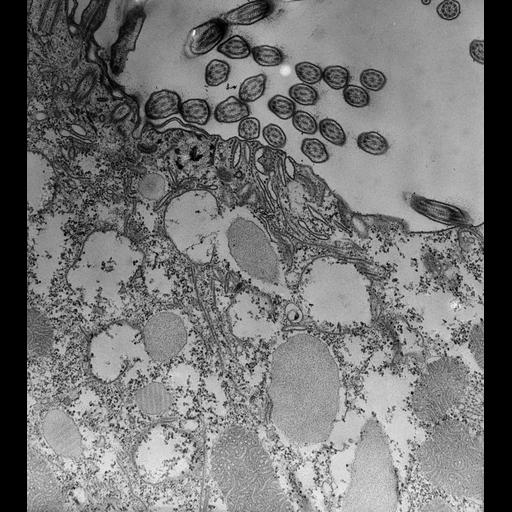
This micrograph shows that the discoidal vesicles meet the cytopharyngeal membrane edge-on where they fuse. Besides the ribbon of 12 microtubules abutting the membrane there is a second small ribbon of 2 or 3 microtubules lying against the cytopharyngeal membrane on the posterior side of each ribbon. This narrow ribbon does not curve away from the membrane but remains close to the membrane and its termination marks the boundary line between the cytopharynx and the nascent vacuole membrane. TEM taken on 7/27/73 by R. Allen with Hitachi HU11A operating at 75kV. Neg. 12,750X. Published in J. Cell Biol. 63:904-922, 1974. Adapted with permission.

Technical Details

Standard glutaraldehyde fixation followed by osmium tetroxide, dehydrated in alcohol and embedded in an epoxy resin. Microtome sections prepared at approximately 75nm thickness. The raw negative was scanned with an Epson Perfection V750 Pro and this high resolution image is best used for quantitative analysis. Additional information available at (http://www5.pbrc.hawaii.edu/allen/).

Biological Sources
NCBI Organism Classification
Paramecium caudatum
Cell Type
cell by organism
eukaryotic cell
Eukaryotic Protist
Ciliated Protist
Cellular Component
oral apparatus
coated pit
vesicle membrane
Biological Context
Biological Process
digestion
digestive system process
Attribution
Names
Richard Allen (University of Hawaii)
Published
J Cell Biol. 1974 Dec;63(3):904-22.
Published
4373478
Citation
Digital Object Identifier (DOI)
doi:10.7295/W9CIL39168
Archival Resource Key (ARK)
ark:/b7295/w9cil39168
Grouping This image is part of a group.
Imaging
Image Type
transmission electron microscopy (TEM)
illumination by electrons
Image Mode
detection of electrons
film
Parameters Imaged
electron density
Source of Contrast
stain with broad specificity
Visualization Methods
stain with broad specificity
uranyl salt
osmium tetroxide
lead salt
Processing History
recorded image
film
Print from negative scanned for Photoshop.
Data Qualifiers
processed data
Dimensions
Spatial Axis Image Size Pixel Size
X 4524px 1.2nm
Y 5063px 1.2nm