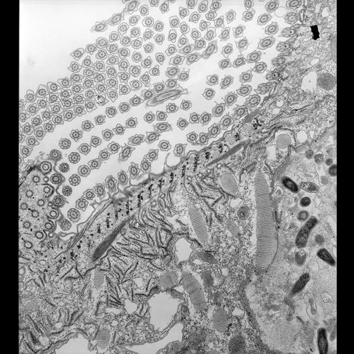
The left edge of the cytopharynx is bordered by a complex architecture designed to transport a pool of flattened vesicles to this edge where they will fuse with the cytopharyngeal membrane to expand this membrane into a nascent phagosome. The buccal cavity itself has the normal pellicle composed of the plasma membrane and alveoli. Internal to this is a system of regularly shaped electron opaque nodes that are bound together by whispy filaments to form a reinforced border. This is part of the filamentous reticulum which is found to a greater or lesser extent underlying the alveolar system of the entire buccal cavity but not the cytopharynx itself. Arising from the nodes are the cytopharyngeal microtubular ribbons that are spaced at regular intervals. These ribbons, composed of about 12 microtubules each, curve into the cytosol over a bundle of filaments called the cytostomal cord which is an extension of the centrin-containing infraciliary lattice. Numerous discoidal vesicles are bound to one side of the microtubules. Associated with the cytosomal cord are regularly spaced tubular extensions of the alveolar sacs that are connected by short fibers to the cord. TEM taken on 3/30/73 by R. Allen with Hitachi HU11A operating at 75kV. Neg. 9,250X. Published in part in J. Cell Biol. 1974. 63:904-22. Adapted with permission.
Standard glutaraldehyde fixation followed by osmium tetroxide, dehydrated in alcohol and embedded in an epoxy resin. Microtome sections prepared at approximately 75nm thickness. The raw negative was scanned with an Epson Perfection V750 Pro and this high resolution image is best used for quantitative analysis. Additional information available at (http://www5.pbrc.hawaii.edu/allen/).
| Spatial Axis | Image Size | Pixel Size |
|---|---|---|
| X | 3616px | 1.1nm |
| Y | 4000px | 1.1nm |