Alternate header for print version


Licensing
Public Domain: This image is in the public domain and thus free of any copyright restrictions. However, as is the norm in scientific publishing and as a matter of courtesy, any user should credit the content provider for any public or private use of this image whenever possible. Learn more
tweet  
*CIL – Cell Image Library accession number. Please use this to reference an image.

CIL:39139*  Cite 
Description

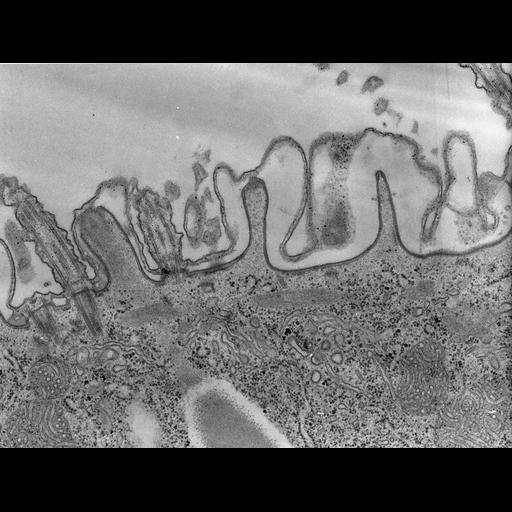
Receptor mediated endocytosis is a process in which extracellular molecules are taken into the cell via coated vesicles that pinch from the plasma membrane. In Paramecium the resulting vesicles fuse with flattened cisterna called early endosomes. Early endosomes accumulate just under the cortex of the cell. Sorting of proteins destined to be returned to the plasma membrane after separating from cargo occurs in the early endosomes and is facilitated by the formation of secondary coated pits and vesicles on the early endosomes that are smaller than the coated and uncoated preendosomal vesicles that arise from the plasma membrane. These small coated pits pinch off and subsequently lose their coats to form carrier vesicles that are roughly 100nm in diameter. These vesicles move deeper into the cell where they bind to microtubular ribbons. TEM taken on 11/4/68 by R. Allen with Philips 300 operating at 60kV. Neg. 20,500X.

Technical Details

Standard glutaraldehyde fixation followed by osmium tetroxide, dehydrated in alcohol and embedded in an epoxy resin. Microtome sections prepared at approximately 75nm thickness. The raw negative was scanned with an Epson Perfection V750 Pro and this high resolution image is best used for quantitative analysis. Additional information available at (http://www5.pbrc.hawaii.edu/allen/).

Biological Sources
NCBI Organism Classification
Paramecium caudatum
Cell Type
cell by organism
eukaryotic cell
Eukaryotic Protist
Ciliated Protist
Cellular Component
coated pit
cell cortex
mitochondrion
glycogen granule
Biological Context
Biological Process
cortical cytoskeleton organization
cytoplasm organization
Attribution
Names
Richard Allen (University of Hawaii)
Citation
Digital Object Identifier (DOI)
doi:10.7295/W9CIL39139
Archival Resource Key (ARK)
ark:/b7295/w9cil39139
Grouping This image is part of a group.
Imaging
Image Type
transmission electron microscopy (TEM)
illumination by electrons
Image Mode
detection of electrons
film
Parameters Imaged
electron density
Source of Contrast
intrinsic mass distribution
stain with broad specificity
Visualization Methods
stain with broad specificity
uranyl salt
gold
Processing History
recorded image
film
Print from negative scanned for Photoshop.
Data Qualifiers
processed data
Sample Preparation
Methods
glutaraldehyde fixed tissue
tissue in vitreous ice embedment
microtome-sectioned tissue
monoclonal antibody
Relation To Intact Cell
microtome-sectioned tissue
Dimensions
Spatial Axis Image Size Pixel Size
X 4708px 1.4nm
Y 3542px 1.4nm