Alternate header for print version


Licensing
Public Domain: This image is in the public domain and thus free of any copyright restrictions. However, as is the norm in scientific publishing and as a matter of courtesy, any user should credit the content provider for any public or private use of this image whenever possible. Learn more
tweet  
*CIL – Cell Image Library accession number. Please use this to reference an image.

CIL:39128*  Cite 
Description

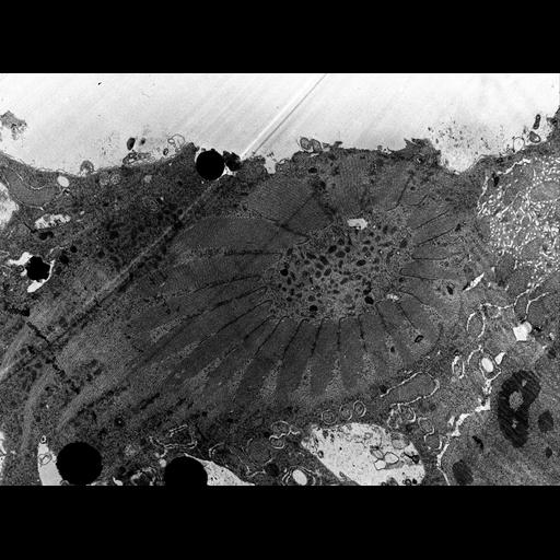
Cytopharyngeal basket in tangential section is composed of a complex of microtubular rods. The lumen of the basket contains many vesicles called cytopharyngeal vesicles. They may provide membrane for the nascent food vacuole, lysosomal and/or lytic enzymes, or all of these. Another role may be to provide vacuolar type ATPases (proton pumps) to the nascent food vacuole. TEM taken on 3/13/69 by R. Allen with Philips 300 operating at 60kV. Neg. 3,570X.

Technical Details

Standard glutaraldehyde fixation followed by osmium tetroxide, dehydrated in alcohol and embedded in an epoxy resin. Microtome sections prepared at approximately 75nm. The raw negative was scanned with an Epson Perfection V750 Pro and this high resolution image is best used for quantitative analysis. Additional information available at (http://www5.pbrc.hawaii.edu/allen/).

Biological Sources
NCBI Organism Classification
Nassula
Cell Type
cell by organism
eukaryotic cell
Eukaryotic Protist
Ciliated Protist
Cell Line
Cambridge Culture Collection, Cambridge, England, catalog # LB1650/1
Cellular Component
oral apparatus
microtubule associated complex
symbiont-containing vacuole
Biological Context
Biological Process
oral apparatus organization
microtubule bundle formation
bacterial symbiont interaction
Attribution
Names
Richard Allen
Citation
Digital Object Identifier (DOI)
doi:10.7295/W9CIL39128
Archival Resource Key (ARK)
ark:/b7295/w9cil39128
Grouping This image is part of a group.
Imaging
Image Type
transmission electron microscopy (TEM)
illumination by electrons
Image Mode
detection of electrons
film
Parameters Imaged
electron density
Source of Contrast
stain with broad specificity
Visualization Methods
stain with broad specificity
osmium tetroxide
uranyl salt
lead salt
Processing History
recorded image
film
unprocessed raw data
Data Qualifiers
raw, unprocessed data
suitable for spatial measurements
Dimensions
Spatial Axis Image Size Pixel Size
X 4000px 1.4nm
Y 2948px 1.4nm