Alternate header for print version


Licensing
Attribution Non-Commercial; No Derivatives:This image is licensed under a Creative Commons Attribution, Non-Commercial, No Derivatives License. View License Deed | View Legal Code
tweet  
*CIL – Cell Image Library accession number. Please use this to reference an image.

CIL:39105*  Cite 
Description

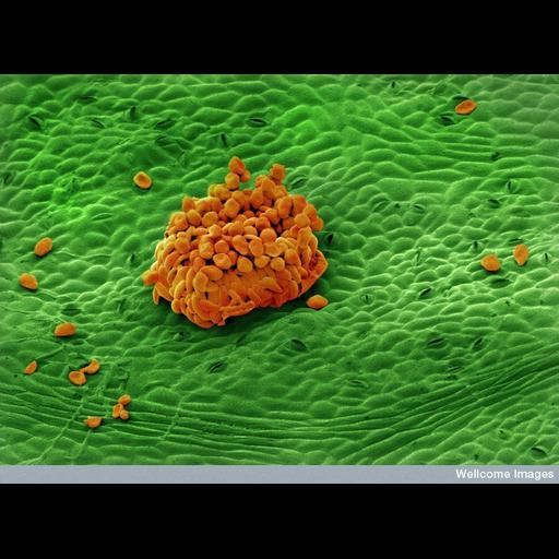
Colorized scanning electron micrograph of rust on a rose leaf. Rose rust is a disease specific to roses and is caused by the parasitic fungus Phragmidium tuberculatum and some other closely related species. This disease occurs during spring and persists until the leaves fall. This images shows rose rust and tiny pores in the leaf (called stomata) visible on the surface of the leaf. Stomata are essential for plant gas exchange. The main group of spores from the fungus is 200uM

Technical Details

B0007248 Rust on rose leaf. Wellcome Images available under the following creative commons usage http://creativecommons.org/licenses/by-nc-nd/2.0/uk/

Biological Sources
NCBI Organism Classification
Phragmidium tuberculatum
Rosa
Cell Type
epidermal cell
fungal spore
Cellular Component
stomata
Biological Context
Biological Process
respiratory gaseous exchange
response to fungus
Attribution
Names
Annie Cavanagh
Link
Wellcome Images
Citation
Digital Object Identifier (DOI)
doi:10.7295/W9CIL39105
Archival Resource Key (ARK)
ark:/b7295/w9cil39105
Imaging
Image Type
recorded image
Image Mode
scanning electron microscopy (SEM)
Parameters Imaged
elastic scattering of electrons
Source of Contrast
differences in deposition of metal shadow
Visualization Methods
visualization of contiguous regions
Processing History
colorized
Sample Preparation
Relation To Intact Cell
whole mounted tissue
Dimensions
Spatial Axis Image Size Pixel Size
X 786px ——
Y 576px ——