Alternate header for print version


Licensing
Attribution Non-Commercial; No Derivatives:This image is licensed under a Creative Commons Attribution, Non-Commercial, No Derivatives License. View License Deed | View Legal Code
tweet  
*CIL – Cell Image Library accession number. Please use this to reference an image.

CIL:39094*  Cite 
Description

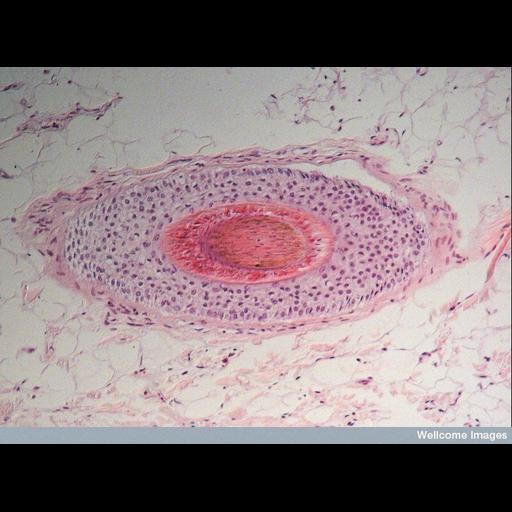
Light micrograph showing a cross section through a hair follicle. The hair shaft is made up of hard dead protein called keratin, which is observed in the center. This is surrounded by skin cells that have been packed together to form the hair follcle. The follicle is surrounded by two layers of cells including erector pili muscle, which causes the hair to stand on 'end'.

Technical Details

B0007314 Cross section of hair follicle. Wellcome Images available under the following creative commons usage http://creativecommons.org/licenses/by-nc-nd/2.0/uk/

Biological Sources
Cell Type
muscle cell
hair follicle cell
Cellular Component
keratin filament
Biological Context
Human Development Anatomy
hair shaft
erector pili muscle
Attribution
Names
Ivor Mason
Link
Wellcome Images
OTHER
Kings College London
Citation
Digital Object Identifier (DOI)
doi:10.7295/W9CIL39094
Archival Resource Key (ARK)
ark:/b7295/w9cil39094
Imaging
Image Type
recorded image
Image Mode
modes of light microscopy
Parameters Imaged
absorption of illumination
Source of Contrast
differences in adsorption or binding of stain
Visualization Methods
hematoxylin
eosin
Processing History
unprocessed raw data
Sample Preparation
Methods
chemically fixed tissue
Relation To Intact Cell
sectioned tissue
Dimensions
Spatial Axis Image Size Pixel Size
X 782px ——
Y 576px ——