Licensing

Attribution_by_nc_nd
Attribution Non-Commercial; No Derivatives:This image is licensed under a Creative Commons Attribution, Non-Commercial, No Derivatives License. View License Deed | View Legal Code
Comments: Powered by Pnyxe

Description

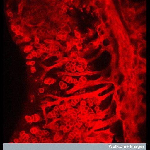
This confocal image shows a 13.5 days post coitum female mouse gonad at high magnification (x40). The developing ovary and mesonephros were fluoresently stained with an endothelial and germ cell marker called PECAM (platelet endothelial cell adhesion molecule). The long projections extending from the right are the endothelial cells migrating into the developing ovary from the adjacent mesonephros. The circular cells are the germ cells in the developing ovary.

Technical Details

B0007505 Germ and endothelial cells. Wellcome Images available under the following creative commons usage http://creativecommons.org/licenses/by-nc-nd/2.0/uk/

Biological Sources

NCBI Organism Classification
Mus musculus
(house mouse)
Cell Type
female germ cell
endothelial cell
Cellular Component
PECAM

Biological Context

Biological Process
cell migration
Mouse Gross Anatomy
TS25,ovary
TS15,mesonephros

Attribution

Name
Dr. Greenfield
Link
Wellcome Images
Other
MRC Harwell

Imaging

Image Type
recorded image
Imaging Mode
confocal microscopy
Parameters Imaged
fluorescence emission
Source of Contrast
distribution of epitope
Visualization Methods
primary antibody plus labeled secondary antibody
Processing History
unprocessed raw data

Sample Preparation

Methods
chemically fixed tissue
Relation To Intact Cell
whole mounted tissue

Dimensions

Spatial Axis Image Size Pixel Size
X 550px ——
Y 556px ——
*CIL – Cell Image Library accession number. Please use this to reference an image.