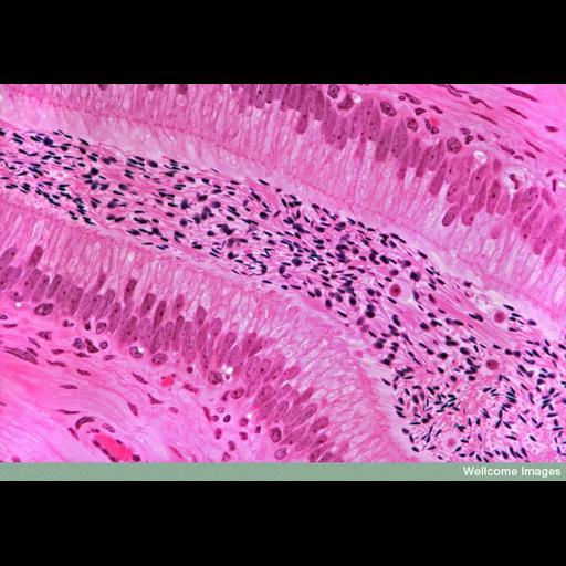
Light micrograph of a longitudinal section of the epididymis. The lining of the duct consists of pseudostratified epithelial cells, which have prominent microvilli on their free surface extending into the lumenal space that contains the spermatozoa. Around the epithelial cells is a layer of smooth muscle, which propels the spermatozoa outwards along the duct during ejaculation. Thousands of spermatozoa are present in the epididymis (the sperm heads are visible as dark spots in the central luminal space) and they are stored here after being produced in the seminiferous tubules of the testis. The cells seen among the spermatozoa are probably lymphocytes. This is a 1.5um (semithin) section stained with haematoxylin and eosin (H+E).
B0007559 Primate epididymis. Wellcome Images available under the following creative commons usage http://creativecommons.org/licenses/by-nc-nd/2.0/uk/
| Spatial Axis | Image Size | Pixel Size |
|---|---|---|
| X | 800px | —— |
| Y | 561px | —— |