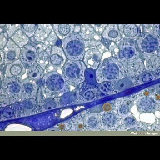
This light micrograph shows the outside edge of two seminiferous tubules of a mouse testis. This section is a 1um thick transverse section, stained with toluidine blue to highlight the cells, with the nucleus staining a darker blue. The dark blue line separating the two seminiferous tubules consists mostly of myoid (muscle) tissue. The majority of cells seen in this image (arranged in layers) are germ cells, which, by repeated cell division, eventually produce spermatozoa. Chromosomes are visible in most of the nuclei of the cells. The cell with a deeply-stained nucleus and even darker nucleolus is a Sertoli cell ('nurse cell') that has fine cytoplasmic extensions branching between the other cells to nurture the developing germ cells.
B0007567 Male germ cells. Wellcome Images available under the following creative commons usage http://creativecommons.org/licenses/by-nc-nd/2.0/uk/
| Spatial Axis | Image Size | Pixel Size |
|---|---|---|
| X | 800px | —— |
| Y | 500px | —— |