Alternate header for print version


Licensing
Attribution Non-Commercial; No Derivatives:This image is licensed under a Creative Commons Attribution, Non-Commercial, No Derivatives License. View License Deed | View Legal Code
tweet  
*CIL – Cell Image Library accession number. Please use this to reference an image.

CIL:39024*  Cite 
Description

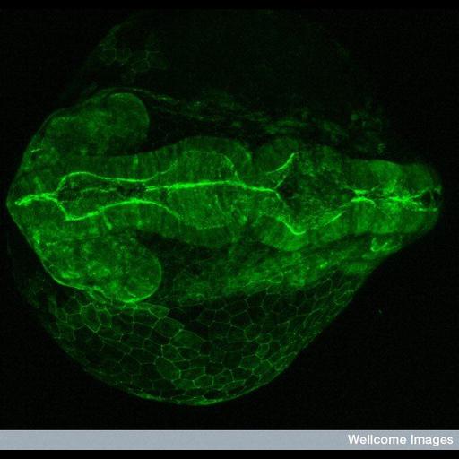
Confocal micrograph of a transgenic zebrafish embryo at 24 hours post-fertilization, showing expression of the fusion protein PARD3_GFP. PARD proteins, which were first identified in C. elegans, are essential for asymmetric cell division and polarized growth. In this image the embryo is viewed from the back (dorsally) sitting on top of a big yolk sac that provides nutrients for its development. GFP expression is seen in the surface of the neuroepithelium and thus highlights the ventricle of the brain, as well as in epithelial cells of the skin recovering the yolk.

Technical Details

B0007774 Dorsal view of an early zebrafish embryo Wellcome Images available under the following creative commons usage http://creativecommons.org/licenses/by-nc-nd/2.0/uk/

Biological Sources
NCBI Organism Classification
Danio rerio
Cell Type
epithelial cell
Cellular Component
PARD3
Biological Context
Biological Process
gene expression
Zebrafish Anatomy
neuroepithelial cell
forebrain ventricle
yolk
Attribution
Names
Monica Folgueira
Steve Wilson
Link
Wellcome Images
Citation
Digital Object Identifier (DOI)
doi:10.7295/W9CIL39024
Archival Resource Key (ARK)
ark:/b7295/w9cil39024
Imaging
Image Type
recorded image
Image Mode
confocal microscopy
Parameters Imaged
fluorescence emission
Source of Contrast
distribution of a specific protein
Visualization Methods
EGFP
Processing History
unprocessed raw data
Sample Preparation
Methods
chemically fixed tissue
Relation To Intact Cell
whole mounted tissue
Dimensions
Spatial Axis Image Size Pixel Size
X 599px ——
X 576px ——