Alternate header for print version


Licensing
Attribution Non-Commercial; No Derivatives:This image is licensed under a Creative Commons Attribution, Non-Commercial, No Derivatives License. View License Deed | View Legal Code
tweet  
*CIL – Cell Image Library accession number. Please use this to reference an image.

CIL:39008*  Cite 
Description

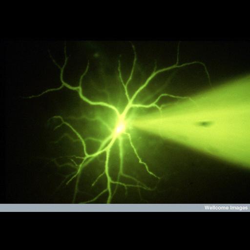
This fluorescent micrograph shows a microelectrode penetrating the cell body of a neurone in the retina of a living eye. The electrode is injecting a fluorescent dye (lucifer yellow) into the cell, filling its branch-like dendrites which form connections with other nerve cells.

Technical Details

B0000061 Injection of retinal cell Wellcome Images available under the following creative commons usage http://creativecommons.org/licenses/by-nc-nd/2.0/uk/

Biological Sources
Cell Type
retinal neuron
Cellular Component
dendrite
neuronal cell body
dendritic branch
Biological Context
Biological Process
dendrite morphogenesis
microinjection
Attribution
Names
David Becker
Link
Wellcome Images
Citation
Digital Object Identifier (DOI)
doi:10.7295/W9CIL39008
Archival Resource Key (ARK)
ark:/b7295/w9cil39008
Imaging
Image Type
recorded image
Image Mode
fluorescence microscopy
Parameters Imaged
fluorescence emission
Source of Contrast
compartmentalization of stain or label
Visualization Methods
lucifer yellow
Processing History
unprocessed raw data
Sample Preparation
Methods
living tissue
Relation To Intact Cell
whole mounted tissue
Dimensions
Spatial Axis Image Size Pixel Size
X 800px ——
Y 559px ——