Alternate header for print version


Licensing
Attribution Non-Commercial; No Derivatives:This image is licensed under a Creative Commons Attribution, Non-Commercial, No Derivatives License. View License Deed | View Legal Code
tweet  
*CIL – Cell Image Library accession number. Please use this to reference an image.

CIL:38996*  Cite 
Description

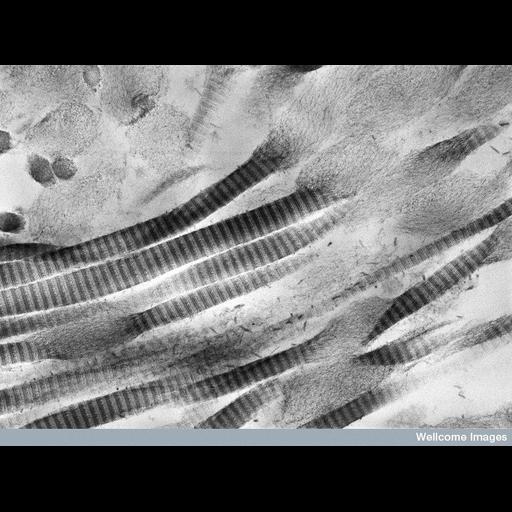
Transmission electron micrograph showing collagen fibrils exhibiting focal swelling and loss of periodic cross banding in the trabecular meshwork of a person with glaucoma. The trabecular meshwork is the structure through which the aqueous humor in the anterior chamber drains into Schlemm's canal. The obstruction of this process may represent a mechanism by which the intraocullar pressure is raised in glaucoma. The proteoglycans that are normally attached to the collagen fibrils have become detached and are seen as small short filaments. The intact fibrils are approximately 120 nm in diameter.

Technical Details

B0001823 Degenerating collagen fibrils in glaucoma. Wellcome Images available under the following creative commons usage http://creativecommons.org/licenses/by-nc-nd/2.0/uk/

Biological Sources
NCBI Organism Classification
Homo sapiens
Cellular Component
extracellular matrix
collagen
Biological Context
Biological Process
collagen fibril organization
Human Development Anatomy
trabecular meshwork
Human Disease
glaucoma
Attribution
Names
Rob Young
Link
Wellcome Images
Citation
Digital Object Identifier (DOI)
doi:10.7295/W9CIL38996
Archival Resource Key (ARK)
ark:/b7295/w9cil38996
Grouping This image is part of a group.
Imaging
Image Type
recorded image
Image Mode
transmission electron microscopy (TEM)
Parameters Imaged
electron density
Source of Contrast
differences in adsorption or binding of stain
Visualization Methods
uranyl salt
cationic dye
Processing History
unprocessed raw data
Sample Preparation
Methods
chemically fixed tissue
Relation To Intact Cell
sectioned tissue
Dimensions
Spatial Axis Image Size Pixel Size
X 774px ——
Y 576px ——