Alternate header for print version


Licensing
Attribution Non-Commercial; No Derivatives:This image is licensed under a Creative Commons Attribution, Non-Commercial, No Derivatives License. View License Deed | View Legal Code
tweet  
*CIL – Cell Image Library accession number. Please use this to reference an image.

CIL:38985*  Cite 
Description

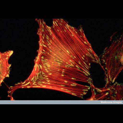
Fluorescent micrograph of rat embryo fibroblast cell growing in serum stained to reveal actin stress fibres (red) and vinculin (component of focal adhesions) in green/yellow.

Technical Details

B0001396 Stress fibres & focal adhesions. Mag. x1000 on 35mm slide. Wellcome Images available under the following creative commons usage http://creativecommons.org/licenses/by-nc-nd/2.0/uk/

Biological Sources
NCBI Organism Classification
Rattus
Cell Type
fibroblast
Cellular Component
stress fiber
focal adhesion
vinculin
Biological Context
Biological Process
cell adhesion
cellular response to starvation
Attribution
Names
Catherine Nobes
Alan Hall
Link
wellcome images
Citation
Digital Object Identifier (DOI)
doi:10.7295/W9CIL38985
Archival Resource Key (ARK)
ark:/b7295/w9cil38985
Imaging
Image Type
recorded image
Image Mode
fluorescence microscopy
Parameters Imaged
fluorescence emission
Source of Contrast
distribution of epitope
Visualization Methods
primary antibody plus labeled secondary antibody
Processing History
unprocessed raw data
Sample Preparation
Methods
chemically fixed tissue
Relation To Intact Cell
dispersed cells in vitro
Dimensions
Spatial Axis Image Size Pixel Size
X 800px ——
Y 557px ——