Alternate header for print version


Licensing
Attribution Non-Commercial; No Derivatives:This image is licensed under a Creative Commons Attribution, Non-Commercial, No Derivatives License. View License Deed | View Legal Code
tweet  
*CIL – Cell Image Library accession number. Please use this to reference an image.

CIL:38969*  Cite 
Description

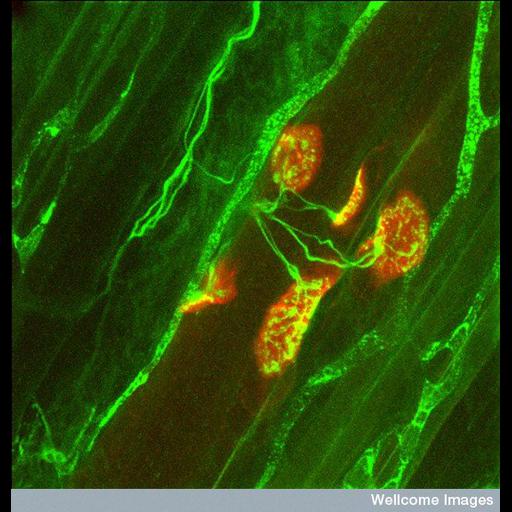
Confocal image of axons (green) making contact with muscle fibers at neuromuscular junctions. As seen here more than one nerve fiber initially contacts each muscle fiber. As development proceeds, the mature innervation pattern is established where only one motor axon contacts each muscle fiber. This transition is called synapse elimination and is thought to involve of competition between the neurons. The neurotransmitter acetylcholine (red) sends the signals from the nerve to makes the muscle contract.

Technical Details

B0004108 Poly-innervated neuromuscular junctions. Wellcome Images available under the following creative commons usage http://creativecommons.org/licenses/by-nc-nd/2.0/uk/

Biological Sources
Cell Type
muscle cell
motor neuron
Cellular Component
synapse
neuromuscular junction
acetylcholine receptor
Biological Context
Biological Process
neuromuscular junction development
Attribution
Names
Tom Gillingwater
Link
Wellcome Images
Citation
Digital Object Identifier (DOI)
doi:10.7295/W9CIL38969
Archival Resource Key (ARK)
ark:/b7295/w9cil38969
Imaging
Image Type
recorded image
Image Mode
confocal microscopy
Parameters Imaged
fluorescence emission
Source of Contrast
distribution of epitope
Processing History
unprocessed raw data
Sample Preparation
Methods
chemically fixed tissue
Relation To Intact Cell
whole
Dimensions
Spatial Axis Image Size Pixel Size
X 550px ——
Y 576px ——