Alternate header for print version


Licensing
Attribution Non-Commercial; No Derivatives:This image is licensed under a Creative Commons Attribution, Non-Commercial, No Derivatives License. View License Deed | View Legal Code
tweet  
*CIL – Cell Image Library accession number. Please use this to reference an image.

CIL:38965*  Cite 
Description

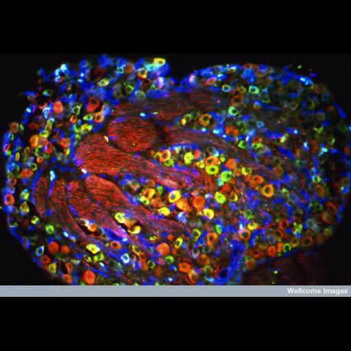
Section through a neonatal dorsal root ganglion (DRG) showing the cell bodies of different populations of sensory nerves. The myelinated A fibers that detect tactile sensations are shown in red. The peptidergic neurons that detect pain and mediate neurogenic inflammation via the release of certain peptides both at the painful site and in the spinal cord are shown in green. The non-peptidergic pain neurons are shown in blue. Bundles of nerve fibers can also be seen within the ganglion.

Technical Details

B0003822 Pain and touch sensory fibers in DRG. Wellcome Images available under the following creative commons usage http://creativecommons.org/licenses/by-nc-nd/2.0/uk/

Biological Sources
Cell Type
sensory neuron
peptidergic neuron
myelinated A fibers
non-peptidergic pain neurons
Cellular Component
neuronal cell body
neuron projection
Biological Context
Biological Process
dorsal root ganglion morphogenesis
Attribution
Names
Simon Beggs
Link
Wellcome Images
Citation
Digital Object Identifier (DOI)
doi:10.7295/W9CIL38965
Archival Resource Key (ARK)
ark:/b7295/w9cil38965
Imaging
Image Type
recorded image
Image Mode
fluorescence microscopy
Parameters Imaged
fluorescence emission
Source of Contrast
differences in adsorption or binding of stain
Visualization Methods
primary antibody plus labeled secondary antibody
Processing History
unprocessed raw data
Sample Preparation
Methods
chemically fixed tissue
Relation To Intact Cell
sectioned tissue
Dimensions
Spatial Axis Image Size Pixel Size
X 800px ——
Y 556px ——