Licensing

Attribution_by_nc_nd
Attribution Non-Commercial; No Derivatives:This image is licensed under a Creative Commons Attribution, Non-Commercial, No Derivatives License. View License Deed | View Legal Code
Comments: Powered by Pnyxe

Description

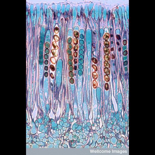
A light micrograph of a thin slice through a cup fungus called Peziza. It grows on decaying wood and organic matter and reproduces itself by producing ascospores. This section shows the fungus' spore containers (asci) each with eight ascospores (shown in brown). As the spores mature, fluid builds up behind them; when the pressure gets too much the tops of the asci open and the spores are flung out into the air. They can cause allergy and irritation in humans including conditions such as dermatitis and hypersensitivity pneumonitis (a condition of the lungs).

Technical Details

B0006300 Ascospores in a Peziza. Wellcome Images available under the following creative commons usage http://creativecommons.org/licenses/by-nc/2.0/uk/

Biological Sources

NCBI Organism Classification
Peziza
Cell Type
ascospore
Cellular Component
ascospore wall

Biological Context

Biological Process
ascospore formation

Attribution

Name
Spike Walker
Link
wellcome images

Imaging

Image Type
recorded image
Imaging Mode
modes of light microscopy
Parameters Imaged
absorption of illumination
Source of Contrast
differences in intrinsic optical density
Visualization Methods
visualization of contiguous regions

Sample Preparation

Methods
chemically fixed tissue
Relation To Intact Cell
sectioned tissue

Dimensions

Spatial Axis Image Size Pixel Size
X 383px ——
Y 576px ——
*CIL – Cell Image Library accession number. Please use this to reference an image.