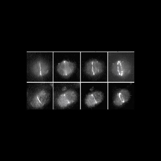
Leaves from wildtype Arabidopsis thaliana were fixed, embedded, sectioned, reacted with antibodies to the FtsZ1-1 protein (upper panels) or FtsZ-2 (lower panels), prokaryotic cell division proteins that are a structural homolog of tubulin. The protein is localized at the site of division which occurs by constricting at the chloroplast mid-point. Shown (from left to right) are focal planes from the bottom, middle and top of the chloroplast, with the projection image at far right revealing the ring-shaped FtsZ distribution.
Leaves were fixed in FAA (formalin acetic acid), embedded in low melting point polyester wax, and 7 micrometer sections cut and attached to poly-L-lysine slides. Sections were de-waxed, treated with an antigen retrieval procedure, and processed for immunofluorescence using incubation with an affinity purified polyclonal antibody, followed by FITC-conjugated secondary antibody. Slides were viewed with an Olympus BH2 microscope equipped with DIC optics. Images were recorded on film, or a video camera (Optronics DEI 750 using a 100x 1.25 NA objective lens. Shown are 3 focal planes. To generate the 3D image shown at far right the z-stack was rotated 30 degrees and projected. See S. Vitha et al. 2001 J Cell Biol 153:111-119.
| Spatial Axis | Image Size | Pixel Size |
|---|---|---|
| X | 342px | —— |
| Y | 184px | —— |