Alternate header for print version


Licensing
Attribution Non-Commercial Share Alike:This image is licensed under a Creative Commons Attribution, Non-Commercial Share Alike License. View License Deed | View Legal Code
tweet  
*CIL – Cell Image Library accession number. Please use this to reference an image.

CIL:38602*  Cite 
Description

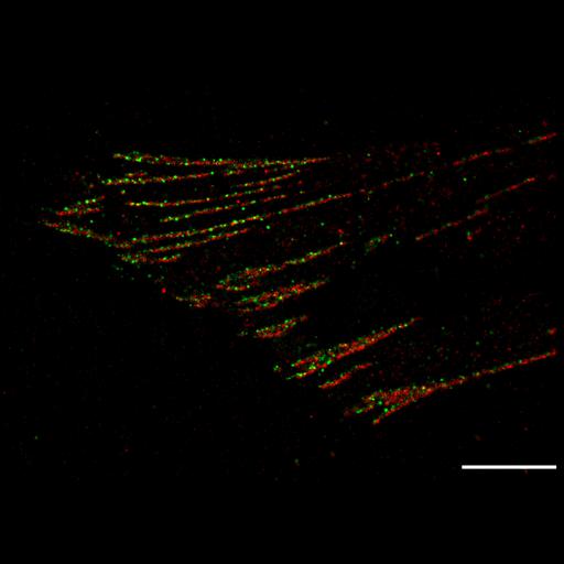
This photoactivation localization microscopy (PALM) image of tdEos-paxillin (green) and PsCFP20-zyxin (red) demonstrates that these two focal adhesion proteins are have very little over-lap when visualized with super-resolution. In contrast, when the same field is visualized with total internal reflection (TIRF) microscopy, the two proteins appear almost completely colocalized CIL 38604. Note the differential interference contrast (DIC) image corresponding to the same image field is also available as CIL 38603. Bar is 2 microns. Image made available by Catherine and James Galbraith and corresponds to Figure 4 in PNAS U S A. 2007 Dec 18;104(51):20308-13.

Biological Sources
NCBI Organism Classification
Homo sapiens
Cell Type
fibroblast
Cell Line
foreskin fibroblast
Cellular Component
focal adhesion
zyxin
paxillin
Biological Context
Biological Process
molecular organization
focal adhesion assembly
Attribution
Names
Hari Shroff
Catherine G Galbraith
James A. Galbraith
Helen White
Jennifer Gillette
Scott Olenych
Michael W. Davidson
Eric Betzig
Published
Proc Natl Acad Sci U S A. 2007 Dec 18;104(51):20308-13. Epub 2007 Dec 12. Erratum in: Proc Natl Acad Sci U S A. 2008 Sep 30;105(39):15220.
Pubmed
18077327
Link
article
Citation
Digital Object Identifier (DOI)
doi:10.7295/W9CIL38602
Archival Resource Key (ARK)
ark:/b7295/w9cil38602
Grouping This image is part of a group.
Imaging
Image Type
portrayed image
Image Mode
PALM (photoactivation localization microscopy)
Parameters Imaged
recorded image
Source of Contrast
fluorescence emission
Visualization Methods
psCFP2
tdEos
Processing History
probability density map
Sample Preparation
Methods
formaldehyde fixed tissue
Relation To Intact Cell
dispersed cells in vitro
Dimensions
Spatial Axis Image Size Pixel Size
X 5788px ——
Y 4091px ——