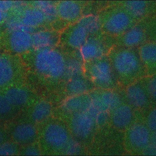
Apoptotic cells produce and transmit the bioactive lipid sphingosine-1-phosphate (S1P) during extrusion. This set of images shows that extrusion is blocked, as well as accumulation of S1P around the dying cell, by SKI II, which blocks conversion of sphingosine to S1P. The apoptotic condensed nucleus is in blue, the actin-myosin ring at the basolateral surface is in red and S1P is in green.
Cultured monolayers were first treated with 30 µM SKI II (EMD) for 10 min. To induce apoptosis, monolayers were exposed to 1,200 µJ/cm2 UV254 irradiation in a UV series II (Spectroline) and incubated for 2 h before fixation. Cells were fixed with 4% formaldehyde in PBS, permeabilized with 0.5% Triton, blocked and incubated with primary antibody for 1 h (50 µg/ml anti-S1P mAb (LPath Inc.)). Secondary antibody was Alexa Fluor 488 goat anti–mouse IgG. Actin was detected with Alexa Fluor 568–phalloidin (Invitrogen). DNA was detected with 5 µM DRAQ5 (Axxora). Confocal micrographs were obtained using an inverted microscope (Eclipse TE300; Nikon) converted for spinning disc confocal microscopy (Andor Technologies) using a 60x Plan Fluor 0.95 oil lens with an electron-multiplied cooled CCD camera 1,000 x 1,000, 8 x 8 mm2 driven by the IQ software (Andor Technologies). ImageJ was used to stack confocal sections into Z series that were then color combined and reconstructed into 3D image using MetaMorph (GE Healthcare). All images were processed further using ImageJ, Photoshop (Adobe), Illustrator (Adobe), and Quicktime Pro (Quicktime) software. This Z-series corresponds to Fig 4D from J Cell Biol. 2011. 193(4): 667-676
| Spatial Axis | Image Size | Pixel Size |
|---|---|---|
| X | 1004px | 1µm |
| Y | 1002px | 1µm |
| Z | 36px | —— |