Alternate header for print version


Licensing
Public Domain: This image is in the public domain and thus free of any copyright restrictions. However, as is the norm in scientific publishing and as a matter of courtesy, any user should credit the content provider for any public or private use of this image whenever possible. Learn more
tweet  
*CIL – Cell Image Library accession number. Please use this to reference an image.

CIL:37339*  Cite 
Description

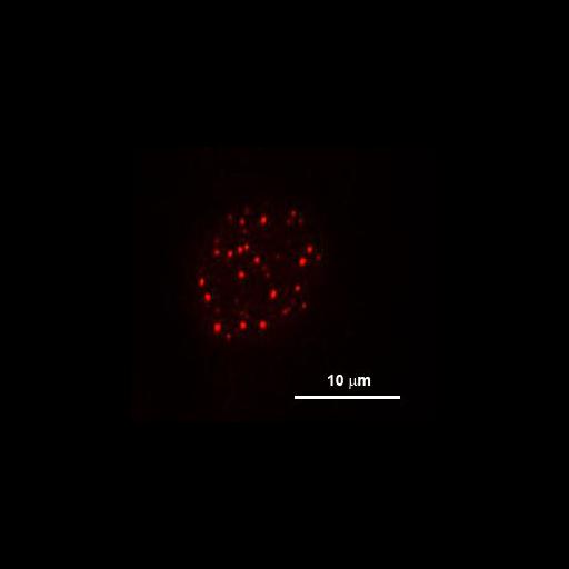
Promyelocytic leukemia nuclear bodies (PML NBs) are dynamic nuclear organelles. They range from 0.1um to 1um in size and exist in most of mammalian cell types. The functions of PML NBs are indicated by their association with a variety of component proteins that are involved in different cellular process. Here shows PML NBs labeled by monoclonal antibody (PML36) in 3T3 cells. .

Technical Details

Cells were rinsed in PBS once and fixed for 15 min in 2% formaldehyde in PBS, pH 7.4. After being washed in PBS for 10 min x3, cells were permeablized 5 min in PBS with 0.2% Triton X-100 and 1% normal goat serum. After being washed in PBS plus 1% goat serum for 10 min x3, the following primary antibody (PML36) were added for 1 h at RT. Cells were rinsed in PBS with 1% normal goat serum for 10 min x3, and secondary Texas Red anti-mouse antibodies were added for 1 h at RT. Cells were rinsed in PBS for 10 min x3 and were observed on a modified Zeiss Axiovert 200M microscope with a plan-neofluar ×40 objective. The microscope was fitted with dual excitation and emission filter wheels and a Photometrics Coolsnap-HQ camera.

Biological Sources
NCBI Organism Classification
Mus musculus
Cell Type
fibroblast
Cell Line
NIH/3T3
Cellular Component
nucleus
PML body
Attribution
Names
Yi-Chun Maria Chen
Citation
Digital Object Identifier (DOI)
doi:10.7295/W9CIL37339
Archival Resource Key (ARK)
ark:/b7295/w9cil37339
Grouping This image is part of a group.
Imaging
Image Type
fluorescence microscopy
Image Mode
fluorescence microscopy
Parameters Imaged
fluorescence emission
Source of Contrast
compartmentalization of stain or label
distribution of epitope
Visualization Methods
TexasRed
primary antibody plus labeled secondary antibody
Processing History
maximum projection
Data Qualifiers
processed data
Sample Preparation
Methods
detergent permeabilized
formaldehyde fixed tissue
Relation To Intact Cell
dispersed cells in vitro
Dimensions
Spatial Axis Image Size Pixel Size
X 274px 0.16µm
Y 248px 0.16µm