Alternate header for print version


Licensing
Attribution Non-Commercial Share Alike:This image is licensed under a Creative Commons Attribution, Non-Commercial Share Alike License. View License Deed | View Legal Code
tweet  
*CIL – Cell Image Library accession number. Please use this to reference an image.

CIL:37317*  Cite 
Description

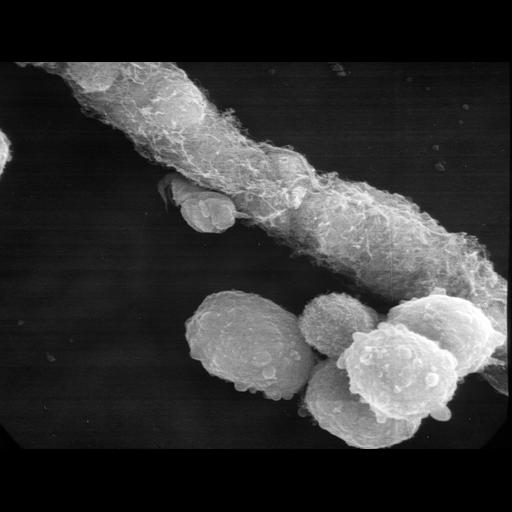
Scanning electron micrograph of single guinea pic pancreatic acinar cells attached to a duct. Image made available by James D. Jamieson and the Department of Cell Biology, Yale University School of Medicine.

Technical Details

iginal 3.25 in. x 4 in. lantern slides were scanned at 600dpi. Original magnification x750.

Biological Sources
NCBI Organism Classification
Cavia porcellus
Cell Type
pancreatic acinar cell
Cellular Component
cell surface
Attribution
Names
James D. Jamieson
A. Amsterdam
Link
George E. Palade EM Slide Collection
Citation
Digital Object Identifier (DOI)
doi:10.7295/W9CIL37317
Archival Resource Key (ARK)
ark:/b7295/w9cil37317
Sample Preparation
Relation To Intact Cell
whole mounted tissue
Dimensions
Spatial Axis Image Size Pixel Size
X 6000px ——
Y 4541px ——