Licensing

Attribution_nc_sa
Attribution Non-Commercial Share Alike:This image is licensed under a Creative Commons Attribution, Non-Commercial Share Alike License. View License Deed | View Legal Code
Comments: Powered by Pnyxe

Description

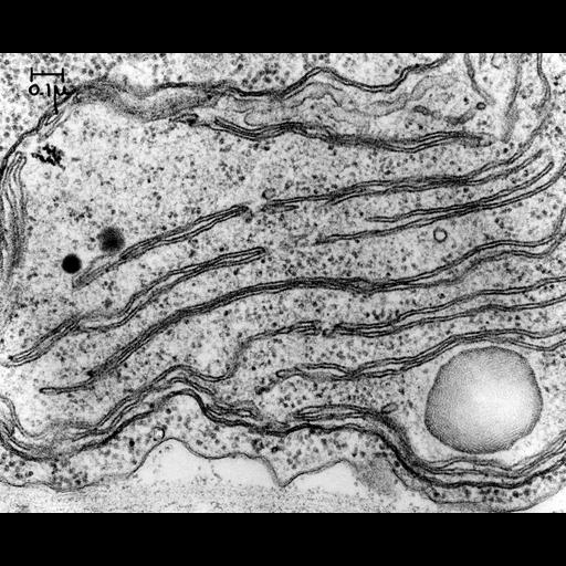
An electron micrograph showing a portion of the single chloroplast of the unicellular green alga Chlamydomonas, an organism extensively used in the Siekvevitz/Palade laboratories at the Rockefeller to study chloroplast membrane biogenesis. These organisms could be grown in large cultures in the dark then, when exposed to light, rapidly and synchronously developed chloroplasts. The characteristic stacked thylakoid membrane pairs are clearly seen. Image made available by James D. Jamieson and the Department of Cell Biology, Yale University School of Medicine.

Technical Details

Original 3.25 in. x 4 in. lantern slides were scanned at 600dpi. Original Magnification: x43,000.

Biological Sources

NCBI Organism Classification
Chlamydomonas reinhardtii
Cellular Component
chloroplast
chloroplast thylakoid membrane

Biological Context

Biological Process
organelle organization
photosynthesis

Attribution

Name
I. Ohad
Link
George E. Palade EM Slide Collection
Date
01/00/1967

Grouping

This image is part of a group.

Imaging

Image Type
recorded image
Imaging Mode
transmission electron microscopy (TEM)
Parameters Imaged
electron density
Source of Contrast
differences in adsorption or binding of stain
Visualization Methods
osmium tetroxide
Processing History
unprocessed raw data

Sample Preparation

Methods
chemically fixed tissue
tissue in plastic embedment
Relation To Intact Cell
sectioned tissue

Dimensions

Spatial Axis Image Size Pixel Size
X 6000px ——
Y 4881px ——
*CIL – Cell Image Library accession number. Please use this to reference an image.