Alternate header for print version


Licensing
Attribution Non-Commercial Share Alike:This image is licensed under a Creative Commons Attribution, Non-Commercial Share Alike License. View License Deed | View Legal Code
tweet  
*CIL – Cell Image Library accession number. Please use this to reference an image.

CIL:37235*  Cite 
Description

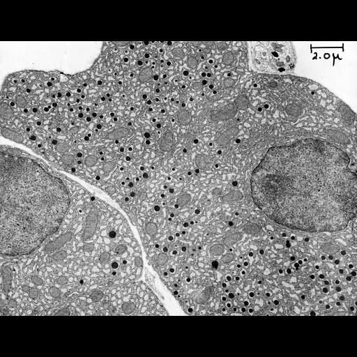
Early transmission electron micrograph showing an alpha cell in the Islet of Langerhans in the guinea pig pancreas. Alpha granules are prominent among the mitochondria, and a portion of nucleus is also present. Image made available by James D. Jamieson and the Department of Cell Biology, Yale University School of Medicine.

Technical Details

Original 3.25 in. x 4 in. lantern slides were scanned at 600dpi. Original Magnification: 3,500.

Biological Sources
NCBI Organism Classification
Cavia porcellus
Cell Type
Langerhans cell
pancreas
Cellular Component
alpha granules
nucleus
mitochondrion
Biological Context
Biological Process
organelle organization
secretion
Attribution
Names
George E. Palade
Link
George E. Palade EM Slide Collection
Citation
Digital Object Identifier (DOI)
doi:10.7295/W9CIL37235
Archival Resource Key (ARK)
ark:/b7295/w9cil37235
Grouping This image is part of a group.
Imaging
Image Type
recorded image
Image Mode
transmission electron microscopy (TEM)
Parameters Imaged
electron density
Source of Contrast
differences in adsorption or binding of stain
Visualization Methods
osmium tetroxide
Processing History
unprocessed raw data
Sample Preparation
Methods
chemically fixed tissue
Relation To Intact Cell
sectioned tissue
Dimensions
Spatial Axis Image Size Pixel Size
X 6000px ——
Y 4619px ——