Licensing

Attribution_nc_sa
Attribution Non-Commercial Share Alike:This image is licensed under a Creative Commons Attribution, Non-Commercial Share Alike License. View License Deed | View Legal Code
Comments: Powered by Pnyxe

Description

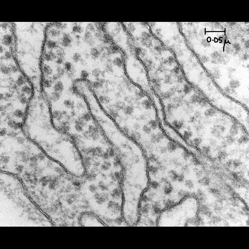
Classic early transmission electron micrograph of a thin section of guinea pig pancreas showing ribosome-studded rough endoplasmic reticulum. Image made available by James D. Jamieson and the Department of Cell Biology, Yale University School of Medicine.

Technical Details

Original 3.25 in. x 4 in. lantern slides were scanned at 600dpi. Original Magnification: 129,000.

Biological Sources

NCBI Organism Classification
Cavia porcellus
(Cavy)
Cell Type
pancreatic cell
Cellular Component
rough endoplasmic reticulum
rough endoplasmic reticulum lumen
rough endoplasmic reticulum membrane
endoplasmic reticulum

Biological Context

Biological Process
organelle organization
protein synthesis
endoplasmic reticulum organization

Attribution

Name
George E. Palade
Link
George E. Palade EM Slide Collection
Date
04/26/1966

Grouping

This image is part of a group.

Imaging

Image Type
recorded image
Imaging Mode
transmission electron microscopy (TEM)
Parameters Imaged
electron density
Source of Contrast
differences in adsorption or binding of stain
Visualization Methods
osmium tetroxide
Processing History
unprocessed raw data

Sample Preparation

Methods
chemically fixed tissue
Relation To Intact Cell
sectioned tissue

Dimensions

Spatial Axis Image Size Pixel Size
X 6000px ——
Y 4942px ——
*CIL – Cell Image Library accession number. Please use this to reference an image.