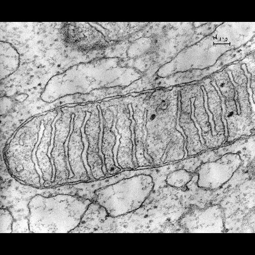
Transmission electron micrograph of mitochondria from guinea pig pancreas. The use of osmium tetroxide as a fixative for electron microscopy was first described by Dr. Palade at the Rockefeller. Another major advance in fixation for electron microscopy was done at Yale by David Sabatini who described the use of glutaraldehyde which not only preserved ultrastructure, but enzymatic activities which opened up the possibility of EM localization of enzymatic function in cells. Image made available by James D. Jamieson and the Department of Cell Biology, Yale University School of Medicine.
References: Palade, G.E. 1952. J. Exp. Med. 95:285-298. Palade, G.E. 1952. Anat. Rec. 114:427-451. Sabatini, D.D, K. Bensch and R.J. Barrnett. 1963. J. Cell. Bio. 17:19-58. Original 3.25 in. x 4 in. lantern slides were scanned at 600dpi. Original magnification 56,000.
| Spatial Axis | Image Size | Pixel Size |
|---|---|---|
| X | 5998px | —— |
| Y | 4975px | —— |