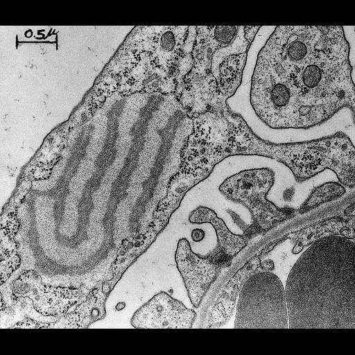
Transmission electron micrograph of a tangential section of a rat glomerulus basement membrane. The series of images in this group have particular historic and cell biologic importance for determining the role of the glomerular basement membrane in urinary filtration, especially the charge properties and size selectivity in retention of proteins in the circulation. The images here clearly show the glomerular filtration barrier. The study also elucidated the role of the basement membrane in some forms of nephrosis. Image made available by James D. Jamieson and the Department of Cell Biology, Yale University School of Medicine.
Relevant citations: Kanwar, Y.S. and M.G. Farquhar. 1979. J. Cell Biol. 81:137-153. Farquhar, M.G., S.L. Wisssig and G.E. Palade. 1961 . J. Exp. Med. 113:47-66. Original 3.25 in. x 4 in. lantern slides were scanned at 600dpi. Original magnification X18,000.
| Spatial Axis | Image Size | Pixel Size |
|---|---|---|
| X | 6000px | —— |
| Y | 5132px | —— |