Licensing

Attribution_nc_sa
Attribution Non-Commercial Share Alike:This image is licensed under a Creative Commons Attribution, Non-Commercial Share Alike License. View License Deed | View Legal Code
Comments: Powered by Pnyxe

Description

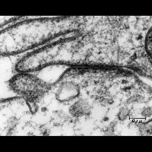
Transmission electron micrograph of tight junction (zonula occludens) between adjacent plasma membranes in the distal kidney tubule. The darkly staining horizontal structure forms the junctional complex. Image made available by James D. Jamieson and the Department of Cell Biology, Yale University School of Medicine.

Technical Details

Original 3.25 in. x 4 in. lantern slides were scanned at 600dpi. Original Magnification: x90,000.

Biological Sources

Cell Type
kidney cell
Cellular Component
tight junction

Biological Context

Biological Process
tight junction assembly
Xenopus Anatomy
distal tubule

Attribution

Name
Marilyn G. Farquhar
Link
George E. Palade EM Slide Collection
Date
01/04/1963

Grouping

This image is part of a group.

Imaging

Image Type
recorded image
Imaging Mode
transmission electron microscopy (TEM)
Parameters Imaged
electron density
Source of Contrast
differences in adsorption or binding of stain
Visualization Methods
osmium tetroxide
Processing History
unprocessed raw data

Sample Preparation

Methods
chemically fixed tissue
Relation To Intact Cell
sectioned tissue

Dimensions

Spatial Axis Image Size Pixel Size
X 6000px ——
Y 4754px ——
*CIL – Cell Image Library accession number. Please use this to reference an image.