Alternate header for print version


Licensing
Attribution Non-Commercial Share Alike:This image is licensed under a Creative Commons Attribution, Non-Commercial Share Alike License. View License Deed | View Legal Code
tweet  
*CIL – Cell Image Library accession number. Please use this to reference an image.

CIL:37131*  Cite 
Description

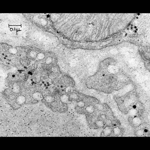
Transmission electron micrograph of rat capillary diaphragm 2h post intravenous administration of ferritin. Ferritin is seen entering plasmalemmal vesicles and into the extracellular space. Image made available by James D. Jamieson and the Department of Cell Biology, Yale University School of Medicine.

Technical Details

Original 3.25 in. x 4 in. lantern slides were scanned at 600dpi. Original magnification X62,000.

Biological Sources
NCBI Organism Classification
Rattus
Cell Type
capillary endothelial cell
Cellular Component
vesicle
Biological Context
Biological Process
regulation of vascular permeability
Attribution
Names
R. Burns
Link
George E. Palade EM Slide Collection
OTHER
10/24/1962
Citation
Digital Object Identifier (DOI)
doi:10.7295/W9CIL37131
Archival Resource Key (ARK)
ark:/b7295/w9cil37131
Grouping This image is part of a group.
Imaging
Image Type
recorded image
Image Mode
transmission electron microscopy (TEM)
Parameters Imaged
electron density
Source of Contrast
differences in adsorption or binding of stain
Visualization Methods
osmium tetroxide
Sample Preparation
Relation To Intact Cell
sectioned tissue
Dimensions
Spatial Axis Image Size Pixel Size
X 6000px ——
Y 4956px ——