Alternate header for print version


Licensing
Attribution Non-Commercial Share Alike:This image is licensed under a Creative Commons Attribution, Non-Commercial Share Alike License. View License Deed | View Legal Code
tweet  
*CIL – Cell Image Library accession number. Please use this to reference an image.

CIL:37127*  Cite 
Description

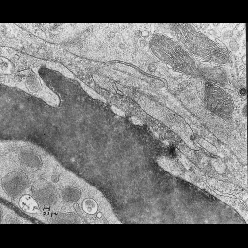
Transmission electron micrograph of mouse capillary intestine fenestrated endothelium with hemoglobin in the lumen lumen. The capillary lumen filled with hemoglobin shows continuities across endothelium. Image made available by James D. Jamieson and the Department of Cell Biology, Yale University School of Medicine.

Technical Details

Reference: Simeonescu, N., M. Simeonescu, and G. E. Palade. 1975. J. Cell Biol. 64:586-607. Original 3.25 in. x 4 in. lantern slides were scanned at 600dpi. Original magnification x14,800

Biological Sources
NCBI Organism Classification
Mus musculus
Cell Type
capillary endothelial cell
fenestrated cell
Biological Context
Biological Process
regulation of vascular permeability
Attribution
Names
George E. Palade
Link
George E. Palde EM Slide Collection
Citation
Digital Object Identifier (DOI)
doi:10.7295/W9CIL37127
Archival Resource Key (ARK)
ark:/b7295/w9cil37127
Grouping This image is part of a group.
Imaging
Image Type
recorded image
Image Mode
transmission electron microscopy (TEM)
Parameters Imaged
electron density
Source of Contrast
differences in adsorption or binding of stain
Visualization Methods
osmium tetroxide
Dimensions
Spatial Axis Image Size Pixel Size
X 6000px ——
Y 4864px ——