Licensing

Copyright
This image is copyright protected. Any public or private use of this image is subject to prevailing copyright laws. Please contact the content provider of this image for permission requests.
Comments: Powered by Pnyxe

Description

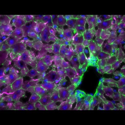
Image is of a section of a liver from a CD-1 mouse treated intravenously with siRNA-Cy5 enapsulated with lipid nanoparticle (LNP201-siRNA-Cy5) for 2 hours. siRNA-Cy5 (red) can be observed in the sinusoids and inside hepatocytes. Cells are outlined with phalloidin-Alexa488 (green) and nuclei are stained with DAPI (blue). The black section in the lower right corner is a vein of the liver. This image is Fig 2F of J Histochem Cytochem 2011 59: 727 and Cover Image for the Journal of Histochemistry and Cytochemistry, August 2011.

Technical Details

7 micron thick cryosection of liver was fixed with 4% paraformaldehyde in PBS for 10 min. Sections were stained with phalloidin-Alexa 488 and DAPI. Images were captured with an Olympus BX61 microscope with a 1.4 mega-pixel progressive scan camera.

Biological Sources

NCBI Organism Classification
Mus musculus
(house mouse)
Cell Type
hepatocyte
sinusoids
Cellular Component
filamentous actin
nucleus

Attribution

Names
Bin Shi
Ed Keough
Andrea Matter
Karen Leander
Stephanie Young
Ed Carlini
Alan B. Sachs
Weikang Tao
Marc Abrams
Bonnie Howell
Laura Sepp-Lorenzino
Published
J Histochem Cytochem 2011 59: 727
Link
http://jhc.sagepub.com/content/59/8/72...
Pubmed
21804077
Data Qualifiers
processed data

Sample Preparation

Methods
formaldehyde fixed tissue
Relation To Intact Cell
cryostat-sectioned tissue

Dimensions

Spatial Axis Image Size Pixel Size
X 1344px ——
Y 1024px ——
*CIL – Cell Image Library accession number. Please use this to reference an image.