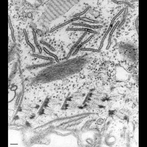
Detail of the origin of the cytopharyngeal microtubules along the left side of the cytopharynx that arise within the filamentous reticulum in contact with its electron-opaque nodes. This system does not arise from the basal bodies of the quadrulus although microtubules do arise along the dorsal edge of the basal bodies of this membranelle and curve between the ends of the cytopharyngeal ribbons and the alveoli of the buccal cavity. The end of the alveoli marks the transition of the buccal cavity into the cytopharynx. As shown on the left of this figure the cytopharyngeal microtubules curve over the cytostomal cord. From here the ribbons fan out into the cytosol. They bind to discoidal vesicles, acidosomes and carrier vesicles and move these vesicles toward the cytopharynx. TEM taken on 3/30/73 by R. Allen with Hitachi HU11A operating at 75kV. Neg. 37,500X. Bar = 0.1µm.
Standard glutaraldehyde fixation followed by osmium tetroxide, dehydrated in alcohol and embedded in an epoxy resin. Microtome sections prepared at approximately 75nm thickness. The negative was printed to paper and the image was scanned to Photoshop. This digitized image is available for qualitative analysis. There is a high resolution version of this image in the library (CIL:39174) which is available for quantitative analysis. Additional information available at (http://www5.pbrc.hawaii.edu/allen/).
| Spatial Axis | Image Size | Pixel Size |
|---|---|---|
| X | 3096px | —— |
| Y | 3481px | —— |