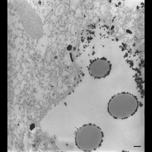
Acid phosphatase is present in tubules retrieved from late DV-III. The tubules and rounded expansions arising as a result of late DV-III membrane retrieval contain an electron-opaque reaction product when the cells are incubated in Gomori’s reagent and lead salts. This indicates that acid phosphatase (AcPase) is being retrieved from the late phagolysosomes before the spent vacuole contents are released at the cytoproct. In fact, spent vacuoles no longer contain active AcPase and are free of AcPase-reaction product before they are defecated. (For details of lysosome recycling see Allen and Fok, J. Cell Biol. 99:1955-1959, 1984). TEM taken on 4/27/84 by R. Allen with Zeiss 10A operating at 80kV. Neg. 19,800X. Bar = 0.2µm. Part published in J. Cell Biol. 99:1955-1959, 1984. Adapted with permission.
Glutaraldehyde fixation followed by Gomori staining, dehydrated in alcohol and embedded in an epoxy resin. Microtome sections prepared at approximately 75nm thickness. The negative was printed to paper and the image was scanned to Photoshop. This image is suitable for qualitative analysis. A high resolution version of this image in the library (CIL:40566) is available for quantitative analysis. Additional information available at (http://www5.pbrc.hawaii.edu/allen/).