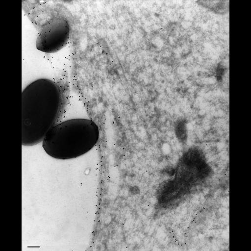
A mAb, B25-1-3, was raised to antigen Q2 (arbitrary code) to the same set of membranes as mAb 132-F4-G6 labels. This mAb was more reactive on frozen sections. To tell the age of vacuoles, cells were exposed to latex beads for 2.5 minutes and then washed free of the beads (0.5 min) and then chased in bead-free buffer for a known time before fixing. In this image the labeled phagosome is a DV-I as it contained 1.1µm beads that were present in the medium for the 3 minutes before the cells were fixed. Some vesicles in the cytosol are also labeled but there is very little nonspecific label over the general cytosol or the contents of the vacuole. TEM taken 10/24/92 by R. Allen with Zeiss 10A operating at 80kV. Neg. 19,800X. Bar = 0.2µm.
To label membranes inside the cell we used very lightly fixed cells (0.25% glutaraldehyde) that were then rapidly frozen in liquid nitrogen and sectioned later at -100oC. These frozen sections were picked up on drops of methylcellulose and transferred to a Formvar-supported grid. The sections were immunogold labeled (15nm gold) to show the location of the specific antigen inside the cell as well as on the cell surface. Microtome sections prepared at approximately 75nm thickness. The negative was printed to paper and the image was scanned to Photoshop. This digitized image is available for qualitative analysis. Additional information available at (http://www5.pbrc.hawaii.edu/allen/).
| Spatial Axis | Image Size | Pixel Size |
|---|---|---|
| X | 2280px | —— |
| Y | 2748px | —— |