Alternate header for print version


Licensing
Public Domain: This image is in the public domain and thus free of any copyright restrictions. However, as is the norm in scientific publishing and as a matter of courtesy, any user should credit the content provider for any public or private use of this image whenever possible. Learn more
tweet  
*CIL – Cell Image Library accession number. Please use this to reference an image.

CIL:36718*  Cite 
Description

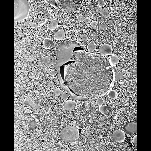
Quick-freeze deep-etch images show a more three dimensional view of acidosomes surrounding a phagosome. Acidosomes are unique in that they contain indentations of their membrane. The lumen has a honeycombed appearance, also unique to these vesicles. Their E-face is almost free of IMPs while their P-face has many large particles which leave pits in the E-face. Acidosomes maintain a close association with the phagosome membrane. In this micrograph the difference between the lumens of pretrichocyst vesicles and acidosomes can be compared. TEM taken on 5/26/92 by R. Allen with Zeiss 10A operating at 80kV. Neg. 9,780X. Bar = 0.5µm. A print of the negative was scanned and processed in Photoshop. This image is best used for qualitative analysis. Additional information available at (http://www5.pbrc.hawaii.edu/allen/).

Biological Sources
NCBI Organism Classification
Paramecium multimicronucleatum
Cell Type
cell by organism
eukaryotic cell
Eukaryotic Protist
Ciliated Protist
Cellular Component
vesicle membrane
early phagosome
trichocyst
Biological Context
Biological Process
vesicle fusion
phagosome acidification
Attribution
Names
Richard Allen (University of Hawaii)
Citation
Digital Object Identifier (DOI)
doi:10.7295/W9CIL36718
Archival Resource Key (ARK)
ark:/b7295/w9cil36718
Imaging
Image Type
transmission electron microscopy (TEM)
illumination by electrons
Image Mode
electron density
Parameters Imaged
elastic scattering of electrons
electron density
elevation
Source of Contrast
differences in deposition of metal shadow
Visualization Methods
transmission electron microscopy (TEM)
Processing History
freeze_fracture/freeze_etch
shadowing and plating
recorded image
film
Print of negative scanned for Photoshop
Data Qualifiers
processed data
Dimensions
Spatial Axis Image Size Pixel Size
X 1940px ——
Y 2400px ——