Alternate header for print version


Licensing
Public Domain: This image is in the public domain and thus free of any copyright restrictions. However, as is the norm in scientific publishing and as a matter of courtesy, any user should credit the content provider for any public or private use of this image whenever possible. Learn more
tweet  
*CIL – Cell Image Library accession number. Please use this to reference an image.

CIL:36701*  Cite 
Description

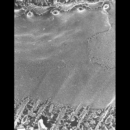
The E-fracture faces of the plasma membrane and cytopharyngeal membranes are continuous but differ in appearance since the cytopharyngeal membrane is studded with IMPs while the plasma membrane has few IMPs. A portion of the plasma membrane is covered by the P-face of the outer alveolar sac membrane which also has many IMPs. The alveolus does not cover the cytopharynx membrane. Four parasomal sacs along the cytopharyngeal edge of the quadrulus are exposed. Details of the attachment of the cytopharyngeal ribbons to the cytopharyngeal membrane are shown including part of five fractured ribbons and the two accessory microtubules lying just posterior to each cytopharyngeal ribbon that are also bound to the membrane. A fiber actually links the ribbon to the membrane. (For details see Allen, J. Cell Biol. 63:904-922, 1974. TEM taken on 5/18/88 by C. Schroeder with Zeiss 10A operating at 80kV. Neg. 40,000X. Bar = 0.1µm.

Biological Sources
NCBI Organism Classification
Paramecium multimicronucleatum
Cell Type
cell by organism
eukaryotic cell
Eukaryotic Protist
Ciliated Protist
Cellular Component
plasma membrane
oral apparatus
microtubule associated complex
Biological Context
Biological Process
oral apparatus organization
Attribution
Names
Richard Allen (University of Hawaii)
C. Schroeder
Citation
Digital Object Identifier (DOI)
doi:10.7295/W9CIL36701
Archival Resource Key (ARK)
ark:/b7295/w9cil36701
Imaging
Image Type
transmission electron microscopy (TEM)
illumination by electrons
Image Mode
electron density
Parameters Imaged
elastic scattering of electrons
electron density
elevation
Source of Contrast
differences in deposition of metal shadow
Visualization Methods
transmission electron microscopy (TEM)
Processing History
freeze_fracture/freeze_etch
shadowing and plating
recorded image
film
Print of negative scanned for Photoshop
Data Qualifiers
processed data
Dimensions
Spatial Axis Image Size Pixel Size
X 2901px ——
Y 3717px ——