Alternate header for print version


Licensing
Public Domain: This image is in the public domain and thus free of any copyright restrictions. However, as is the norm in scientific publishing and as a matter of courtesy, any user should credit the content provider for any public or private use of this image whenever possible. Learn more
tweet  
*CIL – Cell Image Library accession number. Please use this to reference an image.

CIL:36699*  Cite 
Description

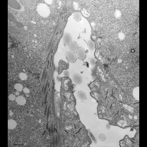
A view of the right side of the cytopharynx showing the groove and its associated microtubules running longitudinally along the groove (for cross sections see Allen, J. Ultrastruct. Res. 63:64-78, 1980, Fig. 4) and bundles of postoral microtubules on its cytosolic side. This cell was fed latex beads and treated with DMSO and then incubated in DAB so that discoidal vesicles appear electron opaque. Some discoidal vesicles can be found along the longitudinally oriented microtubules of the groove. The nascent food vacuole has many acidosomes docked at its surface even at the right side of the cytopharynx. TEM taken on 9/14/82 by R. Allen with Hitachi HU11A operating at 75kV. Neg. 8,500X. Bar = 0.5µm.

Technical Details

Standard glutaraldehyde fixation followed by osmium tetroxide, dehydrated in alcohol and embedded in an epoxy resin. Microtome sections prepared at approximately 75nm thickness. The negative was printed to paper and the image was scanned to Photoshop. This digitized image is available for qualitative analysis. Additional information available at (http://www5.pbrc.hawaii.edu/allen/).

Biological Sources
NCBI Organism Classification
Paramecium multimicronucleatum
Cell Type
cell by organism
eukaryotic cell
Eukaryotic Protist
Ciliated Protist
Cellular Component
oral apparatus
food vacuole
microtubule associated complex
Attribution
Names
Richard Allen (University of Hawaii)
Citation
Digital Object Identifier (DOI)
doi:10.7295/W9CIL36699
Archival Resource Key (ARK)
ark:/b7295/w9cil36699
Imaging
Image Type
transmission electron microscopy (TEM)
illumination by electrons
Image Mode
detection of electrons
film
Parameters Imaged
electron density
Source of Contrast
stain with broad specificity
Visualization Methods
stain with broad specificity
osmium tetroxide
uranyl salt
lead salt
Processing History
recorded image
film
Print from negative scanned for Photoshop.
Data Qualifiers
processed data
Dimensions
Spatial Axis Image Size Pixel Size
X 3096px ——
Y 3464px ——