Alternate header for print version


Licensing
Public Domain: This image is in the public domain and thus free of any copyright restrictions. However, as is the norm in scientific publishing and as a matter of courtesy, any user should credit the content provider for any public or private use of this image whenever possible. Learn more
tweet  
*CIL – Cell Image Library accession number. Please use this to reference an image.

CIL:36681*  Cite 
Description

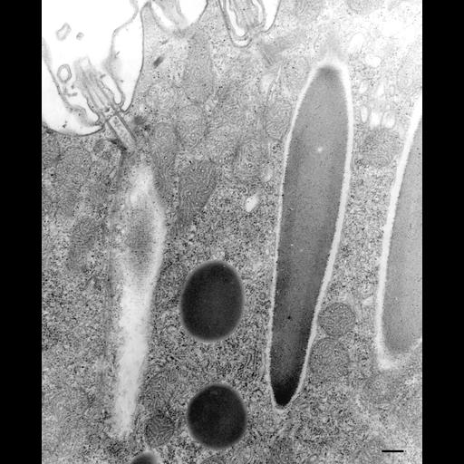
Fully formed trichocysts are moved from the cytosol to the cell surface along microtubules. In this image a bundle of microtubules arising from the proximal end of the basal body lies against the trichocyst membrane along its entire length. The membrane, in grazing section, and the electron-transparent zone are seen. Transport appears to occur tip first along this bundle of microtubules and moves the trichocyst in a direction toward the origin of the microtubular bundle. TEM taken on 4/1/83 by R. Allen with Hitachi HU11A operating at 75kV. Neg. 14,750X. Bar = 0.25µm.

Technical Details

Standard glutaraldehyde fixation followed by osmium tetroxide, dehydrated in alcohol and embedded in an epoxy resin. Microtome sections prepared at approximately 75nm thickness. The negative was printed to paper and the image was scanned to Photoshop. This digitized image is available for qualitative analysis. Additional information available at (http://www5.pbrc.hawaii.edu/allen/).

Biological Sources
NCBI Organism Classification
Paramecium multimicronucleatum
Cell Type
cell by organism
eukaryotic cell
Eukaryotic Protist
Ciliated Protist
Cellular Component
trichocyst
microtubule associated complex
cell cortex
Biological Context
Biological Process
organelle localization
cortical cytoskeleton organization
Attribution
Names
Richard Allen (University of Hawaii)
Citation
Digital Object Identifier (DOI)
doi:10.7295/W9CIL36681
Archival Resource Key (ARK)
ark:/b7295/w9cil36681
Imaging
Image Type
transmission electron microscopy (TEM)
illumination by electrons
Image Mode
detection of electrons
film
Parameters Imaged
electron density
Source of Contrast
stain with broad specificity
Visualization Methods
stain with broad specificity
uranyl salt
lead salt
osmium tetroxide
Processing History
recorded image
film
Print from negative scanned for Photoshop.
Data Qualifiers
processed data
Dimensions
Spatial Axis Image Size Pixel Size
X 2148px ——
Y 2616px ——