Alternate header for print version


Licensing
Public Domain: This image is in the public domain and thus free of any copyright restrictions. However, as is the norm in scientific publishing and as a matter of courtesy, any user should credit the content provider for any public or private use of this image whenever possible. Learn more
tweet  
*CIL – Cell Image Library accession number. Please use this to reference an image.

CIL:36652*  Cite 
Technical Details

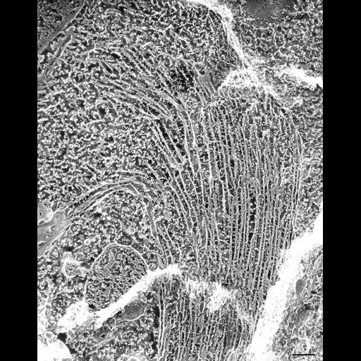
QF-DE of the separation spindle of a micronucleus apparently near a daughter micronucleus (not in section plane). The spindle is filled with microtubules linked together by bridges or by motor proteins. An intact nuclear envelope with pores encloses the spindle. TEM taken on 5/12/91 by R. Allen with Zeiss 10A operating at 80kV. Neg. 19,800X. Bar = 0.25µm.

Biological Sources
NCBI Organism Classification
Paramecium multimicronucleatum
Cell Type
cell by organism
eukaryotic cell
Eukaryotic Protist
Ciliated Protist
Cellular Component
micronucleus
microtubule
Biological Context
Biological Process
nuclear division
Attribution
Names
Richard Allen (University of Hawaii)
Citation
Digital Object Identifier (DOI)
doi:10.7295/W9CIL36652
Archival Resource Key (ARK)
ark:/b7295/w9cil36652
Imaging
Image Type
transmission electron microscopy (TEM)
illumination by electrons
Image Mode
electron density
Parameters Imaged
elastic scattering of electrons
electron density
elevation
Source of Contrast
differences in deposition of metal shadow
Visualization Methods
transmission electron microscopy (TEM)
Processing History
freeze_fracture/freeze_etch
shadowing and plating
recorded image
film
Print of negative scanned for Photoshop
Data Qualifiers
processed data
Dimensions
Spatial Axis Image Size Pixel Size
X 3032px ——
Y 3829px ——