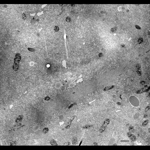
Except that the nuclear envelope remains intact, micronuclear mitosis occurs in a normal way passing through the four stages of prophase, metaphase, anaphase and telophase. In this example a micronucleus was sectioned in early anaphase and shows condensed daughter chromosomes at each pole. Chromosomes are separated by a large bundle of microtubules making up an intranuclear spindle. The separation spindle will become a very long thin tube (see Fig.20 in Allen, Ueno and Fok, J. Protozool. 35:400-407, 1988). The image shown here has very low contrast because the cell was taken from a culture in which a 15 minute treatment in ammonium chloride was used to see what effect this base would have on its acidic compartments. The cell was then incubated in diaminobenzidine. As usual the cristae of mitochondria become electron-opaque following this treatment. TEM taken on 6/11/86 by R. Allen with Zeiss 10A operating at 80kV. Neg. 4,000X. Bar = 1µm.
Standard glutaraldehyde fixation followed by osmium tetroxide, dehydrated in alcohol and embedded in an epoxy resin. Microtome sections prepared at approximately 75nm thickness. The negative was printed to paper and the image was scanned to Photoshop. This digitized image is available for qualitative analysis. Additional information available at (http://www5.pbrc.hawaii.edu/allen/).
| Spatial Axis | Image Size | Pixel Size |
|---|---|---|
| X | 3000px | —— |
| Y | 2832px | —— |