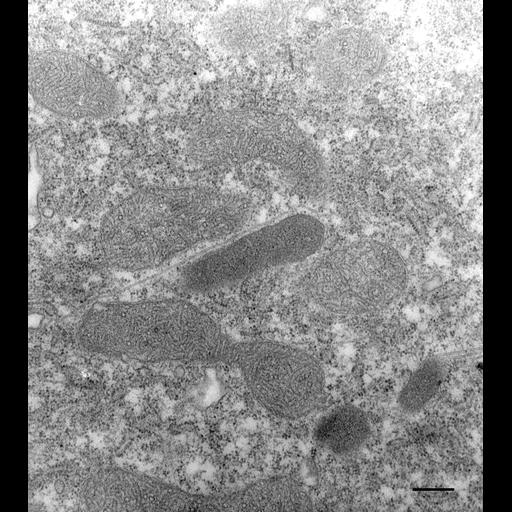
Like other cells Paramecium has peroxisomes. These organelles are usually smaller than mitochondria and have a granular appearance and contain a few tubular-like inclusions. These tubules have not been conclusively shown to be continuous with the single limiting membrane of this organelle. In this case the peroxisome has a microtubule lying along its surface. This cell was exposed to HRP for 30 seconds and then chased in HRP-free medium for 3 minutes. The subsequent incubating with hydrogen peroxidase and diaminobenzidine resulted in an electron opaque reaction product in the peroxisomes not because they took in HRP but because the peroxisomes contain large amounts of catalase that results in the reduction of hydrogen peroxidase. HRP can only enter the living cell by endocytosis, phagocytosis, when trichocysts are discharged, at the open cytoproct or when a CV opens. TEM taken on 6/13/79 by R. Allen with Hitachi HU11A operating at 75kV. Neg. 22,500X. Bar = 0.25µm.
This cell was exposed to HRP for 30 seconds and then chased in HRP-free medium for 3 minutes. The subsequent incubating with hydrogen peroxidase and diaminobenzidine resulted in an electron opaque reaction product in the peroxisomes not because they took in HRP but because the peroxisomes contain large amounts of catalase that results in the reduction of hydrogen peroxidase. This was followed by standard glutaraldehyde fixation followed by osmium tetroxide, dehydrated in alcohol and embedded in an epoxy resin. Microtome sections prepared at approximately 75nm thickness. The negative was printed to paper and the image was scanned to Photoshop. This digitized image is available for qualitative analysis. Additional information available at (http://www5.pbrc.hawaii.edu/allen/).
| Spatial Axis | Image Size | Pixel Size |
|---|---|---|
| X | 2664px | —— |
| Y | 3000px | —— |