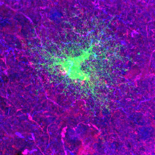
Intracellular dye injections of protoplasmic a 1-week old rat reveals highly ramified spongiform processes that span territories with minimal overlap. Astrocytes were filled with Lucifer Yellow (green) and immunostained to reveal the localization of glial-specific proteins S-100 (red) and GFAP (blue). Optical sections were generated with a single photon confocal microscope. This image has been downsampled from the raw data image which can be accessed using the link provided to the Cell Centered Database.
Male Sprague Dawley rats were anesthetized and perfused transcardially with with Ringer’s solution (37°C), followed by 4% paraformaldehyde (PFA) in PBS (37°C, pH 7.4). Tissue was sectioned (100 µm thick) using a vibratome, and injected with a BX50WI microscope (Olympus, Melville, NY) and, using infrared differential interference contrast videomicroscopy,cells with a small diameter soma (approx. 7–11µm) in the stratum radiatum of CA1 were located and impaled with glass micropipettes filled with either 5% aq. lucifer yellow. The astrocytes were filled using a 0.5 Hz pulses of current until all of the processes were brightly fluorescent. After several astrocytes were filled, a slice was placed in the 4% PFA overnight at 4°C, and subsequently immunostained. Slices were then imaged after coverslipping in Gelvatol. Confocal images were acquired with a BioRad Radiance2000 (Hercules, CA) attached to a Nikon E600FN (Kanagawa, Japan) microscope equipped with a Nikon (Tokyo, Japan) 60× oil objective (NA 1.4). Optical z resolution is 0.25µm. Additional details are provided in Bushong EA, Martone ME, Ellisman MH. Maturation of astrocyte morphology and the establishment of astrocyte domains during postnatal hippocampal development. Int J Dev Neurosci. 2004 Apr;22(2):73-86.
| Spatial Axis | Image Size | Pixel Size |
|---|---|---|
| X | 1052px | —— |
| Y | 1052px | —— |