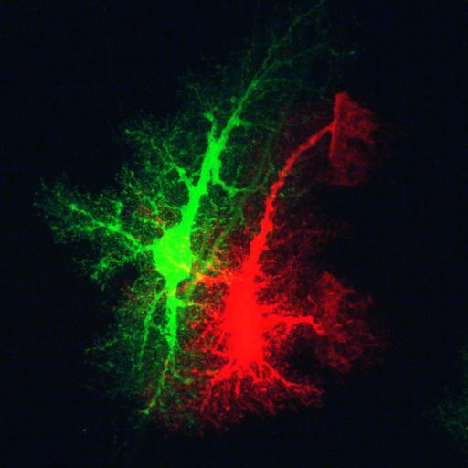
Intracellular dye injections of protoplasmic astrocytes from the CA1 region of hippocampus of 3 week old rat reveals highly ramified spongiform processes that span territories with minimal overlap. Astrocytes were filled with Lucifer Yellow (green) and Alexa 568 (red) Optical sections were generated with a single photon confocal microscope.
Male Sprague Dawley rats, 3 weeks of age, were anaesthetized and perfused transcardially with with Ringer’s solution (37°C), followed by 4% paraformaldehyde (PFA) in PBS (37°C, pH 7.4). Tissue was sectioned (100 µm thick) using a vibratome, and injected with a BX50WI microscope (Olympus, Melville, NY) and, using infrared differential interference contrast videomicroscopy,cells with a small diameter soma (approx. 7–11µm) in the stratum radiatum of CA1 were located and impaled with glass micropipettes filled with either 5% aq. LY, 20mM AlexaFluor 568 in 200mM KCl, or 20mM AlexaFluor 488 in 200mM KCl. The astrocytes were filled using a 0.5 Hz pulses of current until all of the processes were brightly fluorescent. After several astrocytes were filled, a slice was placed in the 4% PFA overnight at 4°C. Slices were then imaged after coverslipping in Gelvatol. Confocal images were acquired with a BioRad Radiance2000 (Hercules, CA) attached to a Nikon E600FN (Kanagawa, Japan) microscope equipped with a Nikon (Tokyo, Japan) 60× oil objective (NA 1.4). Optical resolution of the z-axis, 0.25µm. Additional details are provided in Bushong EA, Martone ME, Ellisman MH. Maturation of astrocyte morphology and the establishment of astrocyte domains during postnatal hippocampal development. Int J Dev Neurosci. 2004 Apr;22(2):73-86.
| Spatial Axis | Image Size | Pixel Size |
|---|---|---|
| X | 1052px | 0.053623µm |
| Y | 1052px | 0.053623µm |