Alternate header for print version


Licensing
Attribution Non-Commercial Share Alike:This image is licensed under a Creative Commons Attribution, Non-Commercial Share Alike License. View License Deed | View Legal Code
tweet  
*CIL – Cell Image Library accession number. Please use this to reference an image.

CIL:36274*  Cite 
Description

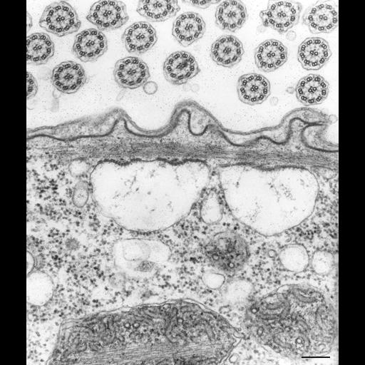
A cross-section and tangential views of three other linkage complexes. The mid piece sits in an indentation of the ER membrane on a bed of 5 slats that pass through the membrane. Rails affix the ER membrane to the myoneme and prevent the vesiculated ER from moving away from the myoneme. Beautifully preserved views of cross sections of cilia. TEM taken on 3/7/72 by R. Allen with Hitachi HU11A operating at 75kV. Neg. 28,500X. Bar = 0.2µm.Published and adapted with permission from J. Cell Biol. 56:559-579, 1973. The negative was printed to paper and the image was scanned to Photoshop. This digitized image is available for qualitative analysis. There is a high resolution version of this image in the library (CIL:39502) which is available for quantitative analysis. Additional information available at (http://www5.pbrc.hawaii.edu/allen/).

Technical Details

Standard glutaraldehyde fixation followed by osmium tetroxide, dehydrated in alcohol and embedded in an epoxy resin. Microtome sections prepared at approximately 75nm thickness. Additional information available at (http://www5.pbrc.hawaii.edu/allen/).

Biological Sources
NCBI Organism Classification
Vorticella convallaria
Cell Type
cell by organism
eukaryotic cell
Eukaryotic Protist
Ciliated Protist
Cell Line
Cambridge Culture Collection, Cambridge, England, # LB1690/1
Cellular Component
pellicle
endoplasmic reticulum
cell cortex
cilium
Attribution
Names
Richard Allen (University of Hawaii)
Published
J Cell Biol. 1973 Feb;56(2):559-79.
Pubmed
4630196
Citation
Digital Object Identifier (DOI)
doi:10.7295/W9CIL36274
Archival Resource Key (ARK)
ark:/b7295/w9cil36274
Grouping This image is part of a group.
Imaging
Image Type
transmission electron microscopy (TEM)
illumination by electrons
Image Mode
detection of electrons
film
Parameters Imaged
electron density
Source of Contrast
stain with broad specificity
Visualization Methods
stain with broad specificity
osmium tetroxide
uranyl salt
lead salt
Processing History
recorded image
film
Print from negative scanned for Photoshop.
Data Qualifiers
processed data
Dimensions
Spatial Axis Image Size Pixel Size
X 2990px ——
Y 3496px ——