Licensing

Attribution_nc_sa
Attribution Non-Commercial Share Alike:This image is licensed under a Creative Commons Attribution, Non-Commercial Share Alike License. View License Deed | View Legal Code
Comments: Powered by Pnyxe

Description

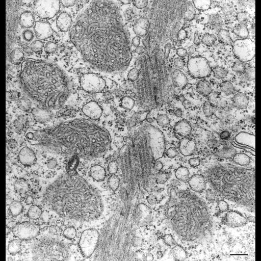
The linkage complex consists of a spindle shaped mid piece bordered, in one plane, by several rails that have subunits that pass through the ER membrane. Thin filaments splay out from both tips of the mid piece and pass toward the surface of the pellicle. The molecular biology or biochemistry of these elements remains to be done. We can only guess that they are involved in calcium movement between the myoneme and the ER and thus in regulating myonemal contraction and relaxation. TEM taken on 4/15/71 by R. Allen with Hitachi HU11A operating at 75kV. Neg. 24,000X. Bar = 0.2µm. Published and adapted with permission from J. Cell Biol. 56:559-579, 1973. The negative was printed to paper and the image was scanned to Photoshop. This digitized image is available for qualitative analysis. There is a high resolution version of this image in the library (CIL:39462) which is available for quantitative analysis. Additional information available at (http://www5.pbrc.hawaii.edu/allen/).

Technical Details

Standard glutaraldehyde fixation followed by osmium tetroxide, dehydrated in alcohol and embedded in an epoxy resin. Microtome sections prepared at approximately 75nm thickness. Additional information available at (http://www5.pbrc.hawaii.edu/allen/).

Biological Sources

NCBI Organism Classification
Vorticella convallaria
Cell Type
cell by organism
eukaryotic cell
Eukaryotic Protist
Ciliated Protist
Cell Line
Cambridge Culture Collection, Cambridge, England, # LB1690/1
Cellular Component
cytoplasm
endoplasmic reticulum
mitochondrion
mitochondrial inner membrane

Biological Context

Biological Process
endoplasmic reticulum organization
mitochondrion organization

Attribution

Name
Richard Allen (University of Hawaii)
Published
J Cell Biol. 1973 Feb;56(2):559-79.
Link
http://www5.pbrc.hawaii.edu/allen/
Pubmed
4630196

Grouping

This image is part of a group.

Imaging

Image Type
transmission electron microscopy (TEM)
illumination by electrons
Imaging Mode
detection of electrons
film
Parameters Imaged
electron density
Source of Contrast
stain with broad specificity
Visualization Methods
stain with broad specificity
osmium tetroxide
uranyl salt
lead salt
Processing History
recorded image
film
Print from negative scanned for Photoshop.
Data Qualifiers
processed data

Dimensions

Spatial Axis Image Size Pixel Size
X 2949px ——
Y 3078px ——
*CIL – Cell Image Library accession number. Please use this to reference an image.