Licensing
Description
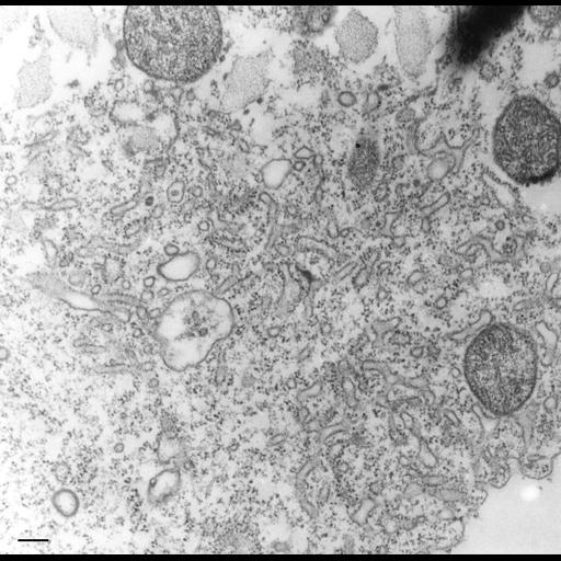
Higher magnification view of the spongiome of the Contractile Vacuole Complex. The spongiome is composed of a three dimensional array of tubules that are continuous with the CV membrane. Many tubules bear peg-like elements on their cytosolic surfaces reminiscent of the decorated tubules of Paramecium. These pegs are probably V-ATPase proton pumps. TEM taken on 5/1/78 by R. Allen with Hitachi HU11A operating at 75kV. Neg. 18,000X. Bar = 0.2µm. The negative was printed to paper and the image was scanned to Photoshop. This digitized image is available for qualitative analysis. There is a high resolution version of this image in the library (CIL:39795) which is available for quantitative analysis. Additional information available at (http://www5.pbrc.hawaii.edu/allen/).
Technical Details
Standard glutaraldehyde fixation followed by osmium tetroxide, dehydrated in alcohol and embedded in an epoxy resin. Microtome sections prepared at approximately 75nm thickness. Additional information available at (http://www5.pbrc.hawaii.edu/allen/).
Biological Sources
- NCBI Organism Classification
- Tetrahymena pyriformis
- (Protozoans)
- Cell Type
- cell by organism
- eukaryotic cell
- Eukaryotic Protist
- Ciliated Protist
- Cellular Component
- contractile vacuole
- contractile vacuolar membrane
- intrinsic to contractile vacuolar membrane
Biological Context
- Biological Process
- contractile vacuole organization
Attribution
- Name
- Richard Allen (University of Hawaii)
- Link
- http://www5.pbrc.hawaii.edu/allen/
Grouping
This image is part of a group.Imaging
- Image Type
- transmission electron microscopy (TEM)
- illumination by electrons
- Imaging Mode
- detection of electrons
- film
- Parameters Imaged
- electron density
- Source of Contrast
- stain with broad specificity
- Visualization Methods
- stain with broad specificity
- osmium tetroxide
- uranyl salt
- lead salt
- Processing History
- recorded image
- film
- Print from negative scanned for Photoshop.
- Data Qualifiers
- processed data
Sample Preparation
- Methods
- glutaraldehyde fixed tissue
- osmium tetroxide fixed tissue
- tissue in epoxy resin embedment
- microtome-sectioned tissue
- Relation To Intact Cell
- microtome-sectioned tissue
Dimensions
| Spatial Axis | Image Size | Pixel Size |
|---|---|---|
| X | 3101px | —— |
| Y | 3040px | —— |